

The value of quantitative electroencephalography in assessing collateral circulation and prognosis in middle cerebral artery occlusive cerebral infarction
-
摘要:
目的 分析脑梗死患者脑电图(EEG)指数的变化,探讨定量脑电图对大脑中动脉闭塞性脑梗死侧支循环及预后的评估价值。 方法 选择2020年6月—2022年12月在丽水市人民医院神经内科就诊,经影像证实为大脑中动脉闭塞性脑梗死患者78例。在发病72 h内完成EEG、多时相计算机体层血管成像(mCTA)和美国国立卫生院卒中量表(NIHSS)评分,在发病后3个月完成改良Barthel指数量表(MBI)评分。侧支循环评估采用mCTA ASITN/SIR侧支循环评估系统。通过Pearson法对EEG指数与mCTA ASITN/SIR评分、NIHSS评分及MBI评分进行相关性分析。 结果 EEG指数δ/α功率比(DAR)、(θ+δ)/(α+β)功率比(DTABR)、配对衍生脑对称指数(pdBSI)与ASITN/SIR评分(r=-0.734、-0.747、-0.759,P<0.01)、MBI评分(r=-0.802、-0.810、-0.853,P<0.01)均呈负相关关系,与NIHSS评分(r=0.876、0.875、0.813,P<0.01)均呈正相关关系。预后不良组EEG指数DAR(t=6.374, P<0.001)、DTABR(t=6.575, P<0.001)、pdBSI(t=9.171, P<0.001)高于预后良好组。ROC曲线分析显示, EEG指数DAR、DTABR、pdBSI对不良结局发生风险均有较高的预测价值(AUC>0.87)。 结论 定量脑电图能有效评估大脑中动脉闭塞性脑梗死神经损伤严重程度和侧支循环,对于3个月神经功能预后也有较好的预测价值。 Abstract:Objective To analyze the changes of electroencephalogram (EEG) indices in patients with cerebral infarction and to explore the value of quantitative EEG on collateral circulation and prognosis of middle cerebral artery occlusive cerebral infarction. Methods Seventy-eight patients with imaging-confirmed middle cerebral artery occlusive stroke who were seen at the Department of Neurology, Lishui People's Hospital between June 2020 and December 2022 were selected. EEG, multitemporal CTA (mCTA), and NIHSS score were completed within 72 hours of onset. MBI scores were completed 3 months after onset. Collateral circulation was assessed using the mCTA ASITN/SIR-based collateral circulation assessment system. Correlation of EEG indices with mCTA ASITN/SIR-based collateral circulation scores, NIHSS scores, and MBI scores was assessed by Pearson correlation analysis. Results The EEG indices DAR, DTABR and pdBSI were negatively correlated with ASITN/SIR collateral circulation scores (r=-0.734, -0.747, -0.759; P < 0.01), MBI scores (r=-0.802, -0.810, -0.853; P < 0.01) and positively correlated with NIHSS scores (r=0.876, 0.875, 0.813; P < 0.01). The EEG indices DAR (t=6.374, P < 0.001), DTABR (t=6.575, P < 0.001) and pdBSI (t=9.171, P < 0.001) were higher in the poor prognosis group than in the good prognosis group. ROC curve analysis showed that the EEG indices DAR, DTABR and pdBSI all had high predictive value for the risk of patients developing adverse outcomes in patients (AUC>0.87). Conclusion Quantitative EEG can effectively assess the severity of middle cerebral artery occlusive stroke and collateral circulation, and also has a good predictive value for 3-month neurological prognosis. -
Key words:
- Quantitative electroencephalography /
- Ischemic stroke /
- Collateral circulation /
- Assessment /
-

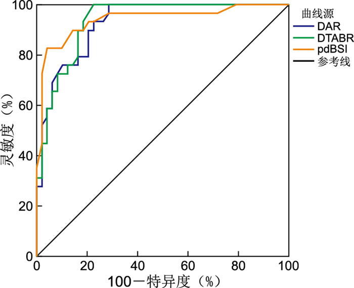
图 1 EEG指数预测AIS不良结局的ROC曲线
Figure 1. ROC curve of the EEG index for predicting adverse outcomes in AIS
表 1 EEG指数与mCTA ASITN/SIR、NIHSS、MBI的相关性
Table 1. Correlation of EEG indices with mCTA ASITN/SIR, NIHSS, and MBI
EEG指数 中位数(区间)a Pearson相关系数(r值)b mCTA ASITN/SIR NIHSS MBI DAR 3.17(0.81~15.21) -0.734 0.876 -0.802 DTABR 2.95(0.78~13.84) -0.747 0.875 -0.810 pdBSI 0.25(0.02~0.67) -0.759 0.813 -0.853 注:a括号内表示最小值到最大值。b均P<0.001。  下载: 导出CSV
表 2 不同预后AIS患者EEG指数比较(x±s)
Table 2. Comparison of EEG index in AIS patients with different prognosis(x±s)
组别 例数 DAR DTABR pdBSI MBI≤60分 29 5.99±2.76 5.52±2.49 0.38±0.10 MBI>60分 49 2.57±1.14 2.32±1.07 0.19±0.08 t值 6.374 6.575 9.171 P值 <0.001 <0.001 <0.001
下载: 导出CSV
表 3 EEG指数对AIS不良结局的预测价值
Table 3. Predictive value of the EEG index for adverse outcomes in AIS
EEG指数 AUC 95% CI SE P值 DAR 0.927 0.873~0.981 0.028 <0.001 DTABR 0.935 0.884~0.985 0.026 <0.001 pdBSI 0.940 0.881~1.000 0.030 <0.001
下载: 导出CSV
-
[1] MENON B K, D'ESTERRE C D, QAZI E M, et al. Multiphase CT angiography: a new tool for the imaging triage of patients with acute ischemic stroke[J]. Radiology, 2015, 275(2): 510-520. doi: 10.1148/radiol.15142256 [2] RABILLER G, HE J W, NISHIJIMA Y, et al. Perturbation of brain oscillations after ischemic stroke: a potential biomarker for post-stroke function and therapy[J]. Int J Mol Sci, 2015, 16(10): 25605-25640. doi: 10.3390/ijms161025605 [3] HUANG H, NIU Z, LIU G, et al. Early consciousness disorder in acute large hemispheric infarction: an analysis based on quantitative EEG and brain network characteristics[J]. Neurocrit Care, 2020, 33(2): 376-388. doi: 10.1007/s12028-020-01051-w [4] SEKER F, PEREIRA-ZIMMERMANN B, PFAFF J, et al. Collateral scores in acute ischemic stroke: a retrospective study assessing the suitability of collateral scores as standalone predictors of clinical outcome[J]. Clin Neuroradiol, 2020, 30(4): 789-793. doi: 10.1007/s00062-019-00858-1 [5] LIEBESKIND D S, SABER H, XIANG B, et al. Collateral circulation in thrombectomy for stroke after 6 to 24 hours in the DAWN trial[J]. Stroke, 2022, 53(3): 742-748. doi: 10.1161/STROKEAHA.121.034471 [6] FINNIGAN S P, ROSE S E, CHALK J B. Rapid EEG changes indicate reperfusion after tissue plasminogen activator injection in acute ischaemic stroke[J]. Clin Neurophysiol, 2006, 117(10): 2338-2339. doi: 10.1016/j.clinph.2006.06.718 [7] FERREIRA L O, MATTOS B G, JÓIA DE MELLO V, et al. Increased relative delta bandpower and delta indices revealed by continuous qEEG monitoring in a rat model of ischemia-reperfusion[J]. Front Neurol, 2021, 12: 645138. DOI: 10.3389/fneur.2021.645138. [8] 刘秀颖, 蓝瑞芳. 急性缺血性脑卒中定量脑电图特征与CT灌注成像参数的相关性[J]. 上海交通大学学报(医学版), 2021, 41(1): 62-65. https://www.cnki.com.cn/Article/CJFDTOTAL-SHEY202101013.htmLIU X Y, LAN R F. Correlation between quantitative electroencephalogram features and CT perfusion imaging parameters in acute ischemic stroke[J]. Journal of Shanghai Jiaotong University(Medical Science), 2021, 41(1): 62-65. https://www.cnki.com.cn/Article/CJFDTOTAL-SHEY202101013.htm [9] FINNIGAN S P, ROSE S E, WALSH M, et al. Correlation of quantitative EEG in acute ischemic stroke with 30-day NIHSS score: comparison with diffusion and perfusion MRI[J]. Stroke, 2004, 35(4): 899-903. doi: 10.1161/01.STR.0000122622.73916.d2 [10] AJČEVIĆ M, FURLANIS G, NACCARATO M, et al. Hyper-acute EEG alterations predict functional and morphological outcomes in thrombolysis-treated ischemic stroke: a wireless EEG study[J]. Med Biol Eng Comput, 2021, 59(1): 121-129. doi: 10.1007/s11517-020-02280-z [11] HU Y, WANG Y, ZHANG R, et al. Assessing stroke rehabilitation degree based on quantitative EEG index and nonlinear parameters[J]. Cogn Neurodyn, 2023, 17(3): 661-669. doi: 10.1007/s11571-022-09849-4 [12] SCHLEIGER E, WONG A, READ S, et al. Poststroke QEEG informs early prognostication of cognitive impairment[J]. Psychophysiology, 2017, 54(2): 301-309. doi: 10.1111/psyp.12785 [13] AGIUS ANASTASI A, FALZON O, CAMILLERI K, et al. Brain symmetry index in healthy and stroke patients for assessment and prognosis[J]. Stroke Res Treat, 2017, 2017: 8276136. DOI: 10.1155/2017/8276136. [14] JIANG M, SU Y, LIU G, et al. Predicting the non-survival outcome of large hemispheric infarction patients via quantitative electroencephalography: superiority to visual electroencephalography and the Glasgow Coma Scale[J]. Neurosci Lett, 2019, 706: 88-92. doi: 10.1016/j.neulet.2019.05.007 [15] BENTES C, PERALTA A R, VIANA P, et al. Quantitative EEG and functional outcome following acute ischemic stroke[J]. Clin Neurophysiol, 2018, 129(8): 1680-1687. doi: 10.1016/j.clinph.2018.05.021 [16] 徐金元, 龚敏操. 重症脑血管病患者脑电图分级情况及其与临床预后的关系[J]. 中华全科医学, 2018, 16(7): 1097-1099, 1157. doi: 10.16766/j.cnki.issn.1674-4152.000302XU J Y, GONG M C. EEG grading in patients with severe cerebrovascular disease and its relationship with clinical prognosis[J]. Chinese Journal of General Practice, 2018, 16(7): 1097-1099, 1157. doi: 10.16766/j.cnki.issn.1674-4152.000302 [17] AMINOV A, ROGERS J M, JOHNSTONE S J, et al. Acute single channel EEG predictors of cognitive function after stroke[J]. PLoS One, 2017, 12(10): e0185841. DOI: 10.1371/journal.pone.0185841. -

 点击查看大图
计量
- 文章访问数: 252
- HTML全文浏览量: 180
- PDF下载量: 9
- 被引次数: 0
