

The value of establishing a model in the differentiation of solid pulmonary nodules based on CT radiomics
-
摘要:
目的 探讨CT影像组学模型对实性肺结节良恶性的诊断效能。 方法 回顾性分析安徽医科大学第二附属医院2019年3月—2022年11月经手术、穿刺或临床证实的231例实性肺结节患者CT影像资料,选择典型的231个肺结节,按病理类型分为良性(98例)和恶性(133例)。采用InferScholar软件分别从二维、三维2个角度对病灶轮廓进行勾画,之后用软件提取影像组学特征,将入组病例以7∶ 3的比例分为训练集和测试集。通过Pearson相关系数、显著性检验、LASSO回归分析方法进行特征筛选。在训练集中分别构建二维、三维影像组学特征模型(模型Ⅰ、模型Ⅱ),用测试集来验证,利用ROC曲线下面积来评价模型的预测效能。 结果 从二维、三维2个角度分别提取出919、1 746个影像组学特征,经过筛选,分别得到12、20个最优影像组学特征,用机器算法构建影像组学模型Ⅰ和模型Ⅱ。训练集中模型Ⅰ的AUC为0.97,模型Ⅱ的AUC为0.98;测试集中模型Ⅰ的AUC、灵敏度、特异度、阳性预测值、阴性预测值和准确率分别为0.94(95% CI:0.87~0.98)、83.9%、89.5%、86.7%、87.2%、87.0%;测试集中模型Ⅱ的AUC、灵敏度、特异度、阳性预测值、阴性预测值和准确率分别为0.97(95% CI:0.94~0.99)、75.9%、97.5%、95.7%、84.8%、88.4%。 结论 基于CT影像组学构建的模型能够较好地预测实性肺结节的良恶性,从三维角度构建的模型Ⅱ的诊断效能优于二维角度构建的模型Ⅰ。 Abstract:Objective To explore the diagnostic efficacy of CT radiomics model for benign and malignant solid pulmonary nodules. Methods CT imaging data of 231 cases of solid pulmonary nodules confirmed by surgery, puncture or clinical diagnosis in our hospital from March 2019 to November 2022 were retrospectively analyzed, select 231 typical pulmonary nodules, and they were divided into benign group (98 cases) and malignant group (133 cases) according to pathological types. Infer Scholar software was used to outline the lesion contour from two and three dimensions respectively. The radiomics features were extracted by software, and the enrolled cases were divided into the training set and the test set in a ratio of 7∶ 3. Features were screened by Pearson correlation coefficient, significance test and LASSO regression analysis. Two dimensional and three dimensional radiomics feature models (model Ⅰ and model Ⅱ) were constructed respectively in the training set, and verified by test set. The area under receiver operating characteristic (ROC) curve (AUC) was used to evaluate the predictive efficiency of the model. Results Total 919 and 1 746 radiomics features were extracted from two dimensions and three dimensions respectively. After screening, 12 and 20 optimal radiomics features were obtained, which were used to construct radiomics model Ⅰ and Ⅱ by machine algorithms. The AUC value of model Ⅰ in the training set was 0.97. The AUC value of model Ⅱ in the training set was 0.98. The AUC value, sensitivity, specificity, positive predictive value, negative predictive value and accuracy of model Ⅰ in the test set were 0.94 (95% CI: 0.87-0.98), 83.9%, 89.5%, 86.7%, 87.2%, 87.0%, respectively. The AUC value, sensitivity, specificity, positive predictive value, negative predictive value and accuracy of model Ⅱ were 0.97 (95% CI: 0.94-0.99), 75.9%, 97.5%, 95.7%, 84.8%, 88.4%, respectively. Conclusion The model based on CT radiomics features can well predict the benign and malignant of solid pulmonary nodules. The diagnostic efficacy of model Ⅱ constructed from three-dimensional perspective is better than that of model Ⅰ constructed from two-dimensional perspective. -
Key words:
- Radiomics /
- Solid pulmonary nodules /
- Differential diagnosis
-

图 1 CT图像上的肺结节轮廓勾画
注:A为原始CT横断面肺窗厚层图像,右肺上叶实性肺结节;B为肺结节勾画示意图,从二维角度对病灶轮廓最大层面进行手动勾画;C~E为肺结节勾画示意图,从三维角度对病灶轮廓进行逐层手动勾画。
Figure 1. Outline of pulmonary nodules on CT images

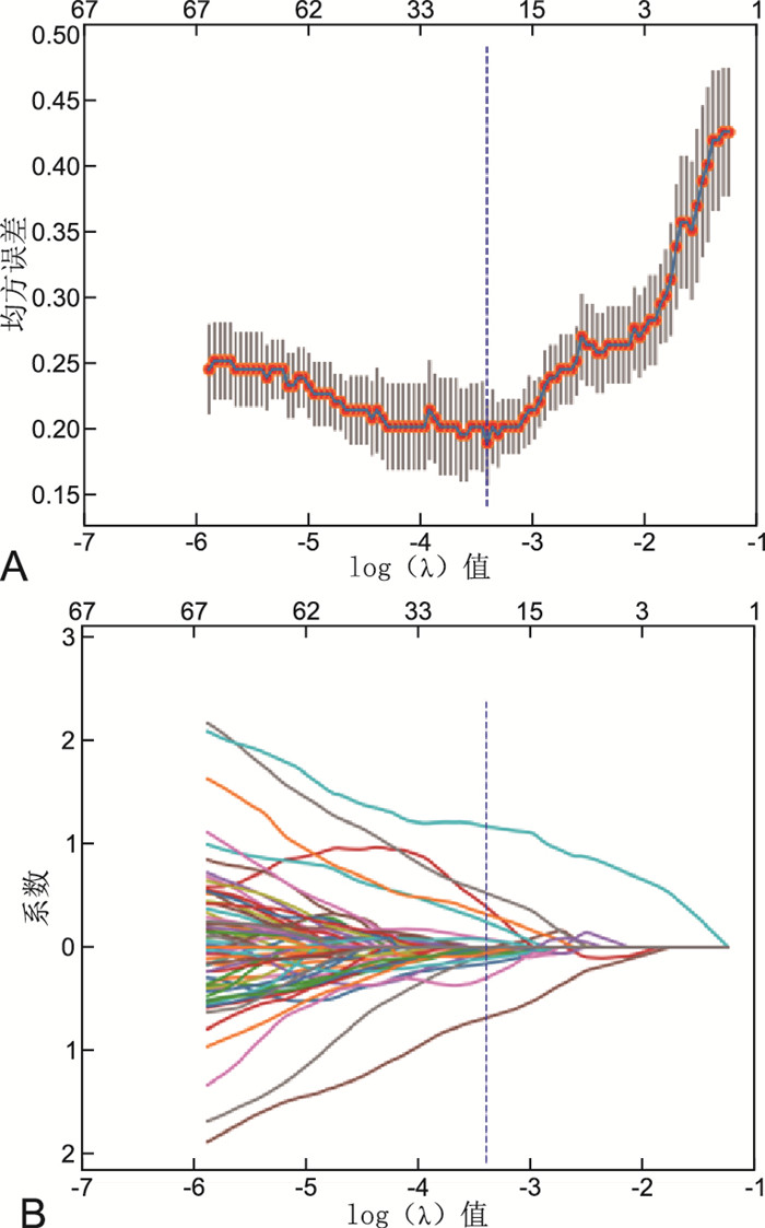
图 2 LASSO回归分析选择影像组学特征(二维)
注:A为通过6-折交叉验证和均方误差筛选出LASSO回归模型中的最优log(λ)值;B中曲线代表最终入选的影像组学特征自变量系数的变化,虚线处对应筛选出的12个影像组学特征。
Figure 2. LASSO regression analysis for selecting radiomics features (2D)

图 3 LASSO回归分析选择影像组学特征(三维)
注:A为通过6-折交叉验证和均方误差筛选出LASSO回归模型中的最优log(λ)值;B中曲线代表最终入选的影像组学特征自变量系数的变化,虚线处对应筛选出的20个影像组学特征。
Figure 3. LASSO regression analysis for selecting radiomics features (3D)

图 4 ROC曲线评估影像组学模型的诊断效能
注:A为模型Ⅰ训练集及测试集预测SPN良恶性的ROC曲线;B为模型Ⅱ训练集及测试集预测SPN良恶性的ROC曲线。
Figure 4. Evaluation of diagnostic efficacy of the radiomics model by ROC curve

图 5 模型Ⅰ及模型Ⅱ测试集的决策曲线
注:A、B分别为模型Ⅰ和模型Ⅱ测试集的决策曲线,体现模型的预测效能。
Figure 5. Decision curves for the test group of model Ⅰ and model Ⅱ test sets
表 1 2种预测模型在训练集与测试集的诊断效能
Table 1. Diagnostic efficiency of two predictive models in training and test sets
模型 灵敏度(%) 特异度(%) 阳性预测值(%) 阴性预测值(%) 准确率(%) AUC 模型Ⅰ 训练集 85.1 94.7 91.9 90.0 90.7 0.97 测试集 83.9 89.5 86.7 87.2 87.0 0.94 模型Ⅱ 训练集 89.9 93.5 91.2 92.6 92.0 0.98 测试集 75.9 97.5 95.7 84.8 88.4 0.97  下载: 导出CSV
-
[1] 犹露, 李邦国. 影像组学在肺结节诊断的研究进展[J]. 中国CT和MRI杂志, 2022, 20(9): 182-184. https://www.cnki.com.cn/Article/CJFDTOTAL-CTMR202209045.htmYOU L, LI B G. Research progress of radiomics in the diagnosis of pulmonary nodules[J]. Chin J CT MRI, 2022, 20(9): 182-184. https://www.cnki.com.cn/Article/CJFDTOTAL-CTMR202209045.htm [2] 郭莹, 高天慧, 赵孟阳, 等. 晚期非小细胞肺癌淋巴细胞亚群及细胞因子与免疫疗效的关系研究[J]. 中华全科医学, 2022, 20(9): 1462-1465. doi: 10.16766/j.cnki.issn.1674-4152.002623GUO Y, GAO T H, ZHAO M Y, et al. Study on the relationship between lymphocyte subsets, cytokines and immune efficacy in advanced non-small cell lung cancer[J]. Chin J Gen Practice, 2022, 20(9): 1462-1465. doi: 10.16766/j.cnki.issn.1674-4152.002623 [3] 余业洲, 赵红, 王龙胜, 等. 基于治疗前CT影像组学评估肺腺癌化疗疗效的研究[J]. 中华全科医学, 2020, 18(4): 623-626, 701. doi: 10.16766/j.cnki.issn.1674-4152.001314YU Y Z, ZHAO H, WANG L S, et al. The study of CT radiomics in assessing the efficacy of pulmonary adenocarcinoma chemotherapy before treatment[J]. Chin J Gen Practice, 2020, 18(4): 623-626, 701. doi: 10.16766/j.cnki.issn.1674-4152.001314 [4] 须民欣, 赵正凯, 杨丹丹, 等. 肺磨玻璃结节的HRCT定量数据与病理性质的相关性分析[J]. 重庆医学, 2021, 50(7): 1178-1181. https://www.cnki.com.cn/Article/CJFDTOTAL-CQYX202107022.htmXU M X, ZHAO Z K, YANG D D, et al. Analysis on correlation between HRCT quantitative data and pathologic properties of pulmonary ground glass nodules[J]. Chongqing Med, 2021, 50(7): 1178-1181. https://www.cnki.com.cn/Article/CJFDTOTAL-CQYX202107022.htm [5] 徐青青, 单文莉, 朱艳, 等. 基于CT影像组学对孤立性肺结节性质分类的预测效能[J]. 南京医科大学学报(自然科学版), 2021, 41(4): 617-623. https://www.cnki.com.cn/Article/CJFDTOTAL-NJYK202104025.htmXU Q Q, SHAN W L, ZHU Y, et al. Predictive performance of the classification of solitary pulmonary nodule based on CT radiomics[J]. J Nanjing Med Univ(Nat Sci), 2021, 41(4): 617-623. https://www.cnki.com.cn/Article/CJFDTOTAL-NJYK202104025.htm [6] PÉREZ-MORALES J, LU H, MU W, et al. Volume doubling time and radiomic features predict tumor behavior of screen-detected lung cancers[J]. Cancer Biomark, 2022, 33(4): 489-501. [7] 周艳, 叶磊, 潘婷婷, 等. 基于灰阶超声的不同影像组学模型鉴别乳腺肿块良恶性的价值[J]. 安徽医学, 2022, 43(2): 127-131. https://www.cnki.com.cn/Article/CJFDTOTAL-AHYX202202001.htmZHOU Y, YE L, PAN T T, et al. The clinical value of different radiomics based on gray scale ultrasound imaging indifferentiating malignant from benign breast masses[J]. Anhui Med J, 2022, 43(2): 127-131. https://www.cnki.com.cn/Article/CJFDTOTAL-AHYX202202001.htm [8] 王晓瑞, 苏晓华, 周全红, 等. CT影像组学鉴别良恶性孤立性肺结节的价值分析[J]. 北京医学, 2022, 44(9): 804-808. https://www.cnki.com.cn/Article/CJFDTOTAL-BJYX202209007.htmWANG X R, SU X H, ZHOU Q H, et al. Differential diagnostic value of CT radiomics in benign and malignant solitary pulmonary nodules[J]. Beijing Med, 2022, 44(9): 804-808. https://www.cnki.com.cn/Article/CJFDTOTAL-BJYX202209007.htm [9] LIU Q, HUANG Y, CHEN H, et al. The development and validation of a radiomic nomogram for the preoperative prediction of lung adenocarcinoma[J]. BMC Cancer, 2020, 20(1): 533. [10] 范卫杰, 张冬. 影像组学及深度学习在肺结节良恶性鉴别诊断中的新理念[J]. 中华肺部疾病杂志(电子版), 2021, 14(5): 549-553. https://www.cnki.com.cn/Article/CJFDTOTAL-ZFBD202105001.htmFAN W J, ZHANG D. New concepts of imaging omics and deep learning in differential diagnosis of benign and malignant pulmonary nodules[J]. Chin J Lung Dis(Electronic Edition), 2021, 14(5): 549-553. https://www.cnki.com.cn/Article/CJFDTOTAL-ZFBD202105001.htm [11] 林天武, 吴佩琪. CT影像组学在孤立性肺结节中的研究进展[J]. 中国医学创新, 2019, 16(21): 168-172. https://www.cnki.com.cn/Article/CJFDTOTAL-ZYCX201921047.htmLIN T W, WU P Q. Advances of Radiomics Research Based on Computed Tomography in Solitary Pulmonary Nodules[J]. Med Innov Chin, 2019, 16(21): 168-172. https://www.cnki.com.cn/Article/CJFDTOTAL-ZYCX201921047.htm [12] 黄栎有, 徐璐, 温林春, 等. CT影像组学模型及深度学习技术预测肺腺癌EGFR突变[J]. 放射学实践, 2022, 37(8): 971-976. https://www.cnki.com.cn/Article/CJFDTOTAL-FSXS202208007.htmHUANG L Y, XU L, WEN L C, et al. Value of CT radiomics models and deep learning technique inpredicting EGFR mutation of lungadeno-carcinoma[J]. Radiol Practice, 2022, 37(8): 971-976. https://www.cnki.com.cn/Article/CJFDTOTAL-FSXS202208007.htm [13] LI H, MENDEL K R, LAN L, et al. Digital mammography in breast cancer: additive value of radiomics of breast parenchyma[J]. Radiology, 2019, 291(1): 15-20. [14] ZHANG Q, PENG Y S, LIU W, et al. Radiomics based on multimodal MRI for the differential diagnosis of benign and malignant breast lesions[J]. J Magn Reson Imaging, 2020, 52(2): 596-607. [15] 杨易, 冯峰, 傅爱燕, 等. 基于MR-T2WI影像组学模型预测宫颈鳞癌临床分期[J]. 中国医学影像技术, 2021, 37(5): 725-730. https://www.cnki.com.cn/Article/CJFDTOTAL-ZYXX202105023.htmYANG Y, FENG F, BO A Y, et al. Radiomics model based on MR-T2WI inpredicting clinical staging of cervical squamous cell carcinoma[J]. Chin J Med Imaging Technol, 2021, 37(5): 725-730. https://www.cnki.com.cn/Article/CJFDTOTAL-ZYXX202105023.htm [16] 崔达华, 赵莹, 刘爱连, 等. 基于动态增强MRI的影像组学列线图预测肝细胞癌切除术后3年复发的价值[J]. 中国临床医学影像杂志, 2019, 30(12): 863-868. https://www.cnki.com.cn/Article/CJFDTOTAL-LYYX201912010.htmCUI D H, ZHAO Y, LIU A L, et al. The value of radiomics nomogram based on dynamic enhanced MRI for predicting the recurrence of HCC in three years after hepatectomy[J]. J Chin Clin Med Imaging, 2019, 30(12): 863-868. https://www.cnki.com.cn/Article/CJFDTOTAL-LYYX201912010.htm [17] 刘畅, 徐有莉, 杨锐, 等. 影像组学评估进展期胰腺癌化疗敏感性及预后[J]. 中国临床医学影像杂志, 2021, 32(2): 120-124. https://www.cnki.com.cn/Article/CJFDTOTAL-LYYX202102014.htmLIU C, XU Y L, YANG R, et al. Radiomics evaluation of chemosensitivity and prognosis of advanced pancreatic cancer[J]. J Chin Clin Med Imaging, 2021, 32(2): 120-124. https://www.cnki.com.cn/Article/CJFDTOTAL-LYYX202102014.htm [18] 孔芳, 段艳华, 宋学林, 等. 高分辨率CT鉴别诊断良、恶性实性孤立性肺结节[J]. 中国医学影像技术, 2021, 37(8): 1168-1172. https://www.cnki.com.cn/Article/CJFDTOTAL-ZYXX202108014.htmKONG F, DUAN Y H, SONG X L, et al. High-resolution CT for differential diagnosis of benign and malignants solitary pulmonary nodules[J]. Chin J Med Imaging Technol, 2021, 37(8): 1168-1172. https://www.cnki.com.cn/Article/CJFDTOTAL-ZYXX202108014.htm [19] MA J C, WANG Q, REN Y C, et al. Automatic lung nodule classification with radiomics approach[C/OL]//PACS and Imaging Informatics: Next Generation and Innovations, 2016[2016-04-05]. https://doi.org/10.1117/12.2220768 .[20] FENG B, CHEN X M, CHEN Y H, et al. Radiomics nomogram for preoperative differentiation of lung tuberculoma from adenocarcinoma in solitary pulmonary solid nodule[J]. Eur J Radiol, 2020, 128: 109022. DOI: 10.1016/j.ejrad.2020.109022. -


 点击查看大图

计量
- 文章访问数: 735
- HTML全文浏览量: 131
- PDF下载量: 30
- 被引次数: 0
