

Establishment and evaluation of risk nomgram model for in-stent restenosis after stent implantation in metaphase and advanced oesophageal carcinoma
-
摘要:
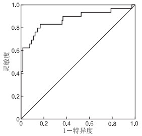
目的 通过分析中晚期食管癌患者支架置入术后支架内再狭窄(in-stent restenosis,ISR)发生的相关影响因素,建立并评估个体化预测中晚期食管癌患者支架置入术后ISR发生风险的列线图模型。 方法 选取2015年5月—2019年6月在瑞安市人民医院消化内科确诊并住院行支架置入术治疗的中晚期食管癌患者160例为研究对象,并根据中晚期食管癌患者支架置入术后ISR的发生与否将研究对象分为术后ISR组(26例)和非术后ISR组(134例)。采用logistic回归模型分析中晚期食管癌患者支架置入术后ISR的独立危险因素。应用列线图在线网站绘制预测中晚期食管癌患者支架置入术后ISR发生风险的列线图模型。采用ROC曲线、校准曲线及Hosmer-Lemeshow拟合优度检验评估列线图模型进行验证。 结果 Logistic回归模型显示,年龄大、合并食管瘘、临床分期为Ⅳ期是中晚期食管癌患者支架置入术后ISR发生的独立危险因素(均P<0.05),支架置入术后接受放射治疗是中晚期食管癌患者支架置入术后ISR发生的保护因素(P<0.05)。ROC曲线结果显示,预测中晚期食管癌患者支架置入术后ISR发生风险的AUC为0.869。校准曲线为斜率接近1的直线,Hosmer-Lemeshow拟合优度检验χ2=5.661,P=0.685。 结论 本研究基于年龄、支架置入术后接受放射治疗、合并食管瘘、临床分期这4项独立影响因素构建的预测中晚期食管癌支架置入术后ISR风险的列线图模型,具有良好的区分度与准确度。 Abstract:Objective To analyse the related factors of in-stent restenosis (ISR) after stent implantation in patients with metaphase and advanced oesophageal carcinoma and to establish and evaluate an individual nomogram model for predicting the risk of ISR. Methods From May 2015 to June 2019, 160 patients with metaphase and advanced oesophageal carcinoma who were diagnosed and hospitalised for stent implantation in the Department of Gastroenterology in Ruian People's Hospital were selected as participants. According to the occurrence of ISR after stent implantation, the patients were divided into post-operative ISR group (n=26) and non-postoperative ISR group (n=134). A logistic regression model was used to analyse the independent risk factors of ISR after stent implantation. A nomogram model for predicting the risk of ISR after stent implantation was developed using nomogram online website. The receiver operating characteristic curve (ROC), calibration curve and Hosmer-Lemeshow goodness-of-fit test were used to evaluate the nomogram model. Results The logistic regression model showed that age, oesophageal fistula and clinical stage were independent risk factors for ISR after stent implantation in patients with metaphase and advanced oesophageal carcinoma (all P < 0.05), and radiotherapy after stent implantation was a protective factor for ISR risk (P < 0.05). ROC results showed that the area under the curve (AUC) for predicting the risk of ISR after stent implantation was 0.869. The calibration curve was a straight line with a slope close to 1, and Hosmer-Lemeshow goodness-of-fit test showed χ2=5.661, P=0.685. Conclusion Based on age, radiotherapy after stent implantation, oesophageal fistula and clinical stage, the nomogram model for predicting the risk of ISR after stent implantation for metaphase and advanced oesophageal carcinoma has good discrimination and accuracy. -
表 1 2组中晚期食管癌患者临床相关指标比较
组别 例数 年龄(x±s,岁) 性别(男/女,例) BMI (x±s) 支架置入术后接受放射治疗[例(%)] 合并食管瘘[例(%)] 病变长度(x±s,cm) 临床分期[例(%)] 病变部位[例(%)] Ⅲ期 Ⅳ期 胸中段 其他 术后ISR组 26 60.32±1.83 16/10 21.91±1.76 7(26.92) 18(69.23) 5.33±1.12 6(23.08) 20(76.92) 17(65.38) 9(34.62) 非术后ISR组 134 59.11±1.95 73/61 22.13±1.59 72(53.73) 49(36.57) 4.95±1.08 72(53.73) 62(46.27) 54(40.30) 80(59.70) 统计量 2.923a 0.440b 0.634a 6.261b 9.545b 1.632a 8.190b 5.552b P值 0.004 0.507 0.527 0.012 0.002 0.105 0.004 0.018 注:a为t值,b为χ2值。  下载: 导出CSV
表 2 中晚期食管癌支架置入术后ISR发生的影响因素分析
项目 B SE Wald χ2 P值 OR值 95%CI 年龄 1.027 0.302 11.561 0.001 2.794 1.545~5.052 支架置入术后接受放射治疗 -2.758 0.924 8.901 0.003 0.063 0.010~0.388 合并食管瘘 3.700 1.146 10.419 0.001 40.460 4.278~382.667 临床分期 2.229 0.909 6.009 0.014 9.288 1.563~55.182
下载: 导出CSV
-
[1] HOLM M O, SKADHAUGE L B, COOK M E, et al. Postoperative nutritional management is insufficient in maintaining body weight in esophageal cancer patients[J]. Clin Nutr, 2020, 40: 563. [2] SAMSON P, PURI V, LOCKHART A C, et al. Adjuvant chemotherapy for patients with pathologic node-positive esophageal cancer after induction chemotherapy is associated with improved survival[J]. J Thorac Cardiovasc Surg, 2018, 156(4): 1725-1735. doi: 10.1016/j.jtcvs.2018.05.100 [3] 王昆仑, 游杰, 郭春旗, 等. 洛铂联合放疗治疗高龄局部晚期食管癌的疗效观察[J]. 现代肿瘤医学, 2019, 27(9): 1532-1535. doi: 10.3969/j.issn.1672-4992.2019.09.016 [4] 唐建萍, 白学松, 裴小玲. 食管支架置入术治疗晚期食管癌的疗效及预后分析[J]. 癌症进展, 2019, 17(7): 791-794. https://www.cnki.com.cn/Article/CJFDTOTAL-AZJZ201907013.htm [5] 龚娇, 孙恒昌, 胡波. 列线图在肿瘤风险预测和预后评估中的应用[J]. 中华检验医学杂志, 2020, 43(6): 614-618. doi: 10.3760/cma.j.cn114452-20200317-00262 [6] TANG J Y, GAO H J, SHI G D, et al. Development and validation of a nomogram prognostic model for patients with neuroendocrine tumors of the thymus[J]. Thorac Cancer, 2020, 11(9): 2457-2464. doi: 10.1111/1759-7714.13556 [7] SAKAI M, SOHDA M, MIYAZAKI T, et al. Association of preoperative nutritional status with prognosis in patients with esophageal cancer undergoing salvage esophagectomy[J]. Anticancer Res, 2018, 38(2): 933-938. [8] 陈国荣, 武源源, 李君艳, 等. 中晚期食管癌患者血浆PAI-1水平及其临床意义[J]. 临床肿瘤学杂志, 2018, 23(4): 335-339. doi: 10.3969/j.issn.1009-0460.2018.04.010 [9] FANG P, MUSALL B C, SON J B, et al. Multimodal imaging of pathologic response to chemoradiation in esophageal cancer[J]. Int J Radiat Oncol Biol Phys, 2018, 102(4): 996-1001. doi: 10.1016/j.ijrobp.2018.02.029 [10] AOYAMA T, ATSUMI Y, HARA K, et al. Postoperative bleeding after esophagectomy for esophageal cancer in patients receiving antiplatelet and anticoagulation treatment[J]. Anticancer Res, 2020, 40(4): 2359-2364. doi: 10.21873/anticanres.14204 [11] 丁瑜, 李伟, 李彬, 等. 光动力疗法与食管支架置入术改善中晚期食管癌所致吞咽困难的对比分析[J]. 中华医学杂志, 2020, 100(5): 378-379, 381. doi: 10.3760/cma.j.issn.0376-2491.2020.05.012 [12] JIN M, RYO K, TAKAAKI S, et al. Clinical significance of palliative esophageal stent placement for esophageal cancer[J]. Gan To Kagaku Ryoho, 2020, 47(3): 487-489. [13] 蒋康, 陆恒, 廖婉玉, 等. 内镜下支架置入治疗食管癌放疗后食管狭窄的疗效观察[J]. 东南国防医药, 2020, 22(2): 200-202. doi: 10.3969/j.issn.1672-271X.2020.02.019 [14] KHAN A, HASHIM Z, NEYAZ Z, et al. Dual airway and esophageal stenting in advanced esophageal cancer with lesions near carina[J]. J Bronchology Interv Pulmonol, 2020, 27(4): 286-293. doi: 10.1097/LBR.0000000000000672 [15] CHEN X, ZHANG W, SUN X, et al. Metabolic syndrome predicts postoperative complications after gastrectomy in gastric cancer patients: Development of an individualized usable nomogram and rating model[J]. Cancer Med, 2020, 9(19): 7116-7124. doi: 10.1002/cam4.3352 [16] 张昊, 谢欣, 周章剑, 等. 列线图预测恶性肿瘤患者PICC导管相关血栓风险的研究[J]. 中国肿瘤临床, 2018, 45(3): 137-141. doi: 10.3969/j.issn.1000-8179.2018.03.877 [17] 王力捷, 沈韦羽, 席勇, 等. 直径≤1 cm肺纯磨玻璃结节中浸润性肺腺癌风险预测的列线图模型研究[J]. 中国医刊, 2019, 54(1): 40-45. doi: 10.3969/j.issn.1008-1070.2019.01.010 [18] 司保才, 刘路光, 郝洪波, 等. 细胞减灭术联合腹腔热灌注化疗治疗腹膜转移结直肠癌预后列线图预测模型研究[J]. 中国实用外科杂志, 2018, 38(1): 105-109. https://www.cnki.com.cn/Article/CJFDTOTAL-ZGWK201801032.htm [19] 刘平, 魏子白, 于俊岩, 等. 中晚期食管癌支架置入术后再狭窄的危险因素分析[J]. 中华消化病与影像杂志(电子版), 2014, 4(2): 58-61. https://www.cnki.com.cn/Article/CJFDTOTAL-ZHYE201402003.htm [20] 胥雄阳, 赵平宗, 蒋丽琳, 等. 覆膜支架置入联合放射治疗在中晚期食管癌治疗中的临床观察[J]. 重庆医学, 2015, 44(25): 3542-3544. doi: 10.3969/j.issn.1671-8348.2015.25.029 -


 点击查看大图

图(3) / 表(2)
计量
- 文章访问数: 358
- HTML全文浏览量: 147
- PDF下载量: 14
- 被引次数: 0
