

Assessment of the effectiveness of multi-slice spiral CT multiparameter for risk stratification of acute pulmonary embolism
-
摘要:
目的 探讨多层螺旋CT(multi-slice spiral CT, MSCT)多参数在急性肺动脉栓塞(acute pulmonary embolism, APE)危险分层中的诊断价值。 方法 纳入2019年1月—2020年10月期间蚌埠医学院第一附属医院经MSCT确诊为APE的74例患者,根据血流动力学状态、右心室功能及心肌是否损伤将患者分为高危组(18例)、中高危组(26例)及中低危组(30例),比较各组间肺动脉阻塞指数(PAOI)及其他影像学参数、临床及实验室指标的差异,用受试者工作特征曲线评价PAOI及心血管参数对于高危和中高危组患者的预测价值,采用多元有序logistic回归分析高危或中高危组APE患者的独立危险因素。 结果 不同组患者在乳酸脱氢酶(LDH)、PAOI、肺动脉干最宽处直径(PTD)、PTD/同一层面升主动脉直径(AAD)、右心室短轴直径(RVD)/左心室短轴直径(LVD)和累及肺段动脉方面差异有统计学意义(均P < 0.001)。PAOI(>38.8%)评估APE高危和中高危患者的灵敏度和特异度分别为65.9%和80.0%,且PAOI值高是APE患者出现高危或中高危的独立危险因素(P < 0.001),RVD/LVD(>1.2)、PTD/AAD(>0.9)评估APE高危及中高危患者的灵敏度和特异度分别为59.1%和93.3%、65.9%和86.7%。 结论 MSCT多参数可能用来评估APE患者的严重程度及预测病情进展,指导临床治疗。 Abstract:Objective To investigate the diagnostic value of multi-slice spiral CT (MSCT) multiparameter in the risk stratification of acute pulmonary embolism (APE). Methods Seventy-four patients diagnosed with APE by MSCT in the First Affiliated Hospital of Bengbu Medical College between January 2019 and October 2020 were included, and the patients were divided into high-risk group (18 patients), medium-high risk group (26 patients) and medium-low risk group (30 patients) according to hemodynamic status, right ventricular function and whether the myocardium was damaged, and the differences in PAOI and other imaging parameters. Clinical and laboratory indices between the groups were compared, and the subject workup characteristic curves were used to evaluate the predictive value of PAOI and cardiovascular. The predictive value of PAOI and cardiovascular parameters for patients in the high-risk and medium-high risk group was evaluated using subject work characteristic curves, and independent risk factors for APE patients in the high-risk or medium-high risk group were analyzed using multivariate ordered logistic regression. Results Statistically significant differences were found in LDH, PAOI, PTD, PTD/AAD, RVD/LVD and involvement of pulmonary segmental arteries in different groups of patients (all P < 0.001). The sensitivity and specificity of PAOI (> 38.8%) for assessing patients at high-risk and medium-high risk of APE were 65.9% and 80.0%, and higher PAOI values are an independent risk factor for patients at high-risk or medium-high risk of APE (P < 0.001). The sensitivity and specificity of RVD/LVD (> 1.2) and PTD/AAD (>0.9) for assessing patients at high-risk and medium-high risk of APE were 59.1% and 93.3%, 65.9% and 86.7%, respectively. Conclusion MSCT multiparameter may be used to assess the severity and predict the progression of APE patients and guide clinical treatment. -

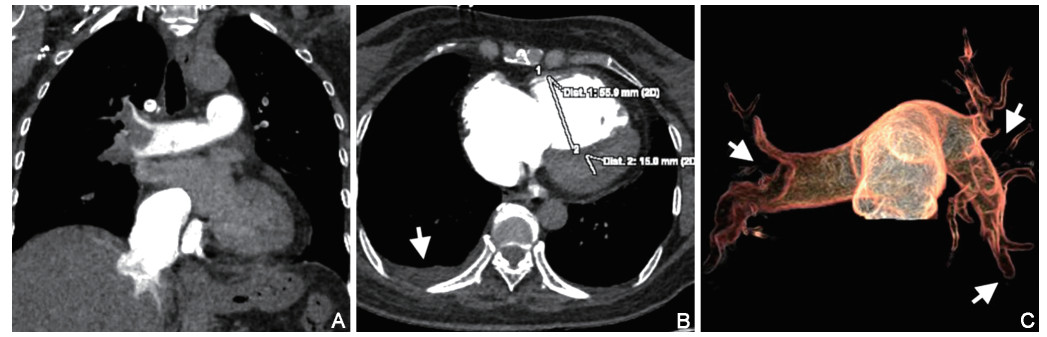
图 1 临床定义为高危APE患者的影像学表现
注:A为冠状位显示右肺动脉干及分支内附壁型血栓;B为RVD、LVD及右侧少量胸腔积液;C为透明血管法显示右肺动脉干、左上肺动脉、左下肺动脉及分支内多发充盈缺损。
表 1 不同临床危险分层APE患者的临床及实验室指标比较
组别 例数 性别[例(%)] 年龄(x±s,岁) 入院时间[M(P25,P75),d] D-D[M(P25,P75),mg/L] CFib(x±s,g/L) AST[M(P25,P75),U/L] LDH[M(P25,P75),U/L] CK/CKMB[M(P25,P75)] HCO3-(x±s,mmol/L) 男性 女性 高危APE 18 7(38.9) 11(61.1) 66.4±11.2 14.0(6.8, 25.8) 18.2(8.0, 58.3) 3.7±1.0 65.0(38.3, 165.0) 938.0(732.3, 1 577.5) 7.1(4.2, 9.4) 23.0±6.2 中高危APE 26 8(30.8) 18(69.2) 65.4±16.6 7.0(1.8, 15.0) 6.7(3.0, 13.3)c 3.4±1.3 32.0(27.8, 47.5)c 546.5(432.8, 651.3)c 3.2(1.8, 6.2)c 24.9±4.1 中低危APE 30 19(63.3) 11(36.7) 65.0±13.8 7.0(3.0, 14.0)c 4.6(1.2, 11.3)c 4.7±1.8ce 36.0(19.0, 52.5)c 599.0(453.8, 599.0)c 3.1(2.1, 6.1)c 25.6±4.9ce 统计量 6.424a 0.061b 8.034d 13.670d 5.514b 10.152d 18.991d 7.966d 6.621b P值 0.040 0.941 0.018 0.001 0.006 0.006 < 0.001 0.019 0.002 注:a为χ2值,b为F值,d为Z值; 与高危组比较,cP<0.05;与中高危组比较,eP<0.05。  下载: 导出CSV
表 2 不同临床危险分层APE患者MSCT影像学指标比较
组别 例数 胸腔(心包)积液[例(%)] PAOI[M(P25,P75),%] PTD[M(P25,P75),mm] PTD/AAD[M(P25,P75)] RVD(x±s, mm) RVD/LVD[M(P25,P75)] 累及肺动脉[例(%)] 累及肺叶动脉[例(%)] 累及肺段动脉[M(P25,P75),n] 高危APE 18 12(66.7) 46.3(25.0, 55.0) 34.5(32.8, 38.0) 1.0(0.9, 1.0) 45.8±7.0 1.3(1.0, 1.8) 14(77.8) 18(100.0) 18.5(10.0, 20.0) 中高危APE 26 12(46.2) 50.0(14.4, 55.0) 30.5(27.0, 33.0)b 0.9(0.8, 0.9) 44.9±7.9 1.4(1.1, 1.6) 19(73.1) 24(92.3) 13.0(5.8, 15.0) 中低危APE 30 18(60.0) 18.8(9.4, 37.5)bc 27.5(26.0, 30.0)b 0.8(0.7, 0.9)b 40.0±6.4bc 1.0(0.9, 1.1)bc 14(46.7)bc 20(66.7)bc 5.5(2.8, 13.0)bc 统计量 2.040a 15.668d 22.041d 20.769d 4.829e 22.013d 6.281a 10.506a 24.744d P值 0.361 <0.001 <0.001 <0.001 0.011 <0.001 0.043 0.003 <0.001 注:a为χ2值, d为Z值, e为F值;与高危组比较,bP<0.05;与中高危组比较,cP<0.05。
下载: 导出CSV
表 3 MSCT影像指标对中高危组APE患者的预测价值
指标 Cut-off值 AUC P值 灵敏度(%) 特异度(%) 95% CI PAOI(%) 38.75 0.770 <0.001 65.9 80.0 0.665~0.876 RVD/LVD 1.24 0.817 <0.001 59.1 93.3 0.723~0.911 PTD/AAD 0.88 0.767 <0.001 65.9 86.7 0.654~0.879 RVD(mm) 41.50 0.715 0.002 79.5 56.7 0.596~0.833 PTD(mm) 30.50 0.746 <0.001 65.9 80.0 0.630~0.862
下载: 导出CSV
表 4 APE患者不同危险分层影响因素的logistic回归分析
变量 B SE Wald χ2 P值 OR值 95% CI PAOI 0.045 0.012 14.088 <0.001 1.046 1.022~1.071 入院时间 0.081 0.028 8.257 0.004 1.084 1.026~1.146 性别 -0.913 0.446 4.198 0.040 0.401 0.167~0.961 HCO3- -0.175 0.051 11.567 0.001 0.840 0.759~0.929 D-D 0.078 0.026 8.914 0.003 1.081 1.027~1.138 注:赋值如下,危险分层为因变量,中低危=0,中高危=1,高危=2。自变量中,性别(男性=0,女性=1);PAOI、入院时间、HCO3-及D-D均以实际值赋值。
下载: 导出CSV
-
[1] FOTI G, SILVA R, FACCIOLI N, et al. Identification of pulmonary embolism: Diagnostic accuracy of venous-phase dual-energy CT in comparison to pulmonary arteries CT angiography[J]. Eur Radiol, 2021, 31(4): 1923-1931. doi: 10.1007/s00330-020-07286-7 [2] YOO H H, NUNES-NOGUEIRA V S, FORTES VILLAS BOAS P J. Anticoagulant treatment for subsegmental pulmonary embolism[J]. Cochrane Database Syst Rev, 2020, 2(2): CD010222. https://pubmed.ncbi.nlm.nih.gov/32030721/ [3] LENFANT M, CHEVALLIER O, COMBY P O, et al. Deep learning versus iterative reconstruction for CT pulmonary angiography in the emergency setting: Improved image quality and reduced radiation dose[J]. Diagnostics (Basel), 2020, 10(8): 558. doi: 10.3390/diagnostics10080558 [4] FU Z, ZHUANG X, HE Y, et al. The diagnostic value of D-dimer with simplified Geneva score (SGS) pre-test in the diagnosis of pulmonary embolism (PE)[J]. J Cardiothorac Surg, 2020, 15(1): 176. doi: 10.1186/s13019-020-01222-y [5] FILOPEI J, BONDARSKY E E, EHRLICH M, et al. Reducing length of stay with the direct oral anti-coagulants in low and intermediate risk pulmonary embolism: A single center experience[J]. J Thromb Thrombolysis, 2020, 50(2): 399-407. doi: 10.1007/s11239-020-02045-3 [6] HIGAZI M M, FATTAH R A R A, ABDELGHANY E A, et al. Efficacy of computed tomography pulmonary angiography as non-invasive imaging biomarker for risk stratification of acute pulmonary embolism[J]. J Clin Imaging Sci, 2020, 10: 49. doi: 10.25259/JCIS_75_2020 [7] KONSTANTINIDES S V, MEYER G, BECATTINI C, et al. 2019 ESC Guidelines for the diagnosis and management of acute pulmonary embolism developed in collaboration with the European Respiratory Society (ERS)[J]. Eur Heart J, 2020, 41(4): 543-603. https://pubmed.ncbi.nlm.nih.gov/31473594/ [8] GUO F, ZHU G, SHEN J, et al. Health risk stratification based on computed tomography pulmonary artery obstruction index for acute pulmonary embolism[J]. Sci Rep, 2018, 8(1): 17897. doi: 10.1038/s41598-018-36115-7 [9] IM D J, HUR J, HAN K, et al. Prognostic value of dual-energy ct-based iodine quantification versus conventional CT in acute pulmonary embolism: A propensity-match analysis[J]. Korean J Radiol, 2020, 21(9): 1095-1103. doi: 10.3348/kjr.2019.0645 [10] 洪都, 徐军, 颜勇卿, 等. Wells评分联合D-二聚体对疑似急性肺栓塞患者的诊断价值[J]. 中华全科医学, 2019, 17(4): 538-542. https://www.cnki.com.cn/Article/CJFDTOTAL-SYQY201904005.htm [11] MINAKAWA M, FUKUDA I, MIYATA H, et al. Outcomes of pulmonary embolectomy for acute pulmonary embolism[J]. Circ J, 2018, 82(8): 2184-2190. doi: 10.1253/circj.CJ-18-0371 [12] EBERLE H, LYN R, KNIGHT T, et al. Clinical update on thrombolytic use in pulmonary embolism: A focus on intermediate-risk patients[J]. Am J Health Syst Pharm, 2018, 75(17): 1275-1285. doi: 10.2146/ajhp170357 [13] PATEL P, PATEL P, BHATT M, et al. Systematic review and meta-analysis of test accuracy for the diagnosis of suspected pulmonary embolism[J]. Blood Adv, 2020, 4(18): 4296-4311. doi: 10.1182/bloodadvances.2019001052 [14] KONSTANTINIDES S, MEYER G. Management of acute pulmonary embolism 2019: What is new in the updated European guidelines?[J]. Intern Emerg Med, 2020, 15(6): 957-966. https://pubmed.ncbi.nlm.nih.gov/32458205/ [15] 陈宗喻, 杜娟, 张先明. 肺血栓栓塞症的临床诊治进展[J]. 中华全科医学, 2020, 18(7): 1181-1184. https://www.cnki.com.cn/Article/CJFDTOTAL-SYQY202007033.htm [16] SHAYGANFAR A, HAJIAHMADI S, ASTARAKI M, et al. The assessment of acute pulmonary embolism severity using CT angiography features[J]. Int J Emerg Med, 2020, 13(1): 15. doi: 10.1186/s12245-020-00272-2 [17] FAGHIHI LANGROUDI T, SHEIKH M, NADERIAN M, et al. The association between the pulmonary arterial obstruction index and atrial size in patients with acute pulmonary embolism[J]. Radiol Res Pract, 2019, 2019: 6025931. https://pubmed.ncbi.nlm.nih.gov/31275649/ [18] LIANG H W, ZHAO D L, LIU X D, et al. ECG-gated pulmonary artery CTA for evaluation of right ventricular function in patients with acute pulmonary embolism[J]. Echocardiography, 2017, 34(2): 257-263. doi: 10.1111/echo.13419 [19] BAX S, JACOB J, AHMED R, et al. Right ventricular to left ventricular ratio at CT pulmonary angiogram predicts mortality in interstitial lung disease[J]. Chest, 2020, 157(1): 89-98. https://pubmed.ncbi.nlm.nih.gov/31351047/ [20] LYHNE M D, SCHULTZ J G, MACMAHON P J, et al. Septal bowing and pulmonary artery diameter on computed tomography pulmonary angiography are associated with short-term outcomes in patients with acute pulmonary embolism[J]. Emerg Radiol, 2019, 26(6): 623-630. doi: 10.1007/s10140-019-01709-9 -

 点击查看大图
图(1) / 表(4)
计量
- 文章访问数: 240
- HTML全文浏览量: 206
- PDF下载量: 4
- 被引次数: 0
