

Analysis of diagnostic valne of stage Ⅰ poorly differentiated lung adenocarcinoma using deep learning model
-
摘要:
目的 肺腺癌高级别成分包括微乳头型、实体型,高级别成分≥20%定义为低分化状态,是患者预后不良的独立预测因素,建议进行肺叶切除。本研究利用4种深度学习模型对低分化腺癌进行预测,比较各模型诊断效能,寻找最佳模型,提高低分化腺癌预测准确性。 方法 回顾性分析西南医科大学附属中医医院2021年10月—2024年3月253个经病理证实的肺腺癌病灶。先对CT图像进行数据预处理及异常数据筛选,然后按照8∶1∶1的比例划分训练验证和测试集,送入到ResNet、MobileNet、DenseNet和EffecientNet 4种模型中,对高级别成分进行预测。 结果 ResNet、MobileNet、DenseNet和EffecientNet四个模型AUC分别为0.757、0.872、0.877、0.812,DenseNet在该任务中展现出色的性能,Accuracy、Precision、Recall和F1-Score分别为84.97%、84.26%、83.28%、84.67%。 结论 4种深度学习模型对肺腺癌高级别成分具有良好的预测作用,DenseNet模型预测准确性更高。 Abstract:Objective Pulmonary adenocarcinoma high-grade components include micropapillary type and solid type, and high-grade components ≥20% are defined as poorly differentiated and are independent predictors of poor prognosis. Lobectomy is recommended. In this study, four kinds of deep learning models were used to predict poorly differentiated adenocarcinoma, and the diagnostic efficiency of each model was compared to find the best model to improve the prediction accuracy of poorly differentiated adenocarcinoma. Methods Retrospective analysis of 253 lung adenocarcinoma lesions confirmed by pathology at the Affiliated Traditional Chinese Medicine Hospital of Southwest Medical University from October 2021 to March 2024. The CT images were preprocessed and abnormal data screened, then the training, validation, and EfficientNet test sets were divided in the ratio of 8∶1∶1 and fed to the four models of ResNet, MobileNet, DenseNet, and EfficientNet for high-level component prediction. Results The AUC values of the four models, ResNet, MobileNet, DenseNet, and EfficientNet, are 0.757, 0.872, 0.877, and 0.812, respectively. DenseNet showed excellent performance in this task. Accuracy, Precision, Recall, and F1-Score were 84.97%, 84.26%, 83.28%, and 84.67%. Conclusion Four kinds of deep learning models have a good predictive effect on high-grade components of lung adenocarcinoma, and the DenseNet model has higher predictive accuracy. -
Key words:
- Lung adenocarcinoma /
- Poorly differentiated /
- Deep learning /
- Model
-

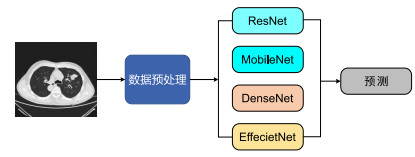
图 1 本研究总体流程图
注:对胸部CT肺窗图像进行预处理后,分别送入4个神经网络模型进行深度学习,再使用多个参数对预测结果进行评估。
Figure 1. The overall flowchart of this study

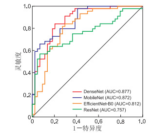
图 2 各模型预测肺腺癌分级状态的ROC曲线
Figure 2. The ROC curves of each model for predicting the grading status of lung adenocarcinoma

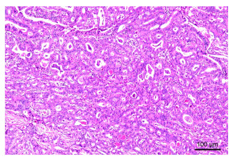
图 3 肺低分化腺癌(实体型)HE染色结果(200×)
Figure 3. HE staining results of lung poorly differentiated adenocarcinoma (solid type) (200×)
表 1 4种模型评估结果
Table 1. The overall flowchart of this study
模型 AUC 准确率(%) 精度(%) 召回率(%) F1分数(%) ResNet 0.757 70.12 69.77 70.24 68.96 MoboleNet 0.872 83.25 84.11 83.87 81.10 DenseNet 0.877 84.97 84.26 83.28 84.67 EfficentNet 0.812 75.87 74.24 77.97 76.03  下载: 导出CSV
-
[1] NICHOLSON A G, TSAO M S, BEASLEY M B, et al. The 2021 WHO classification of lung tumors: impact of advances since 2015[J]. J Thorac Oncol, 2022, 17(3): 362-387. doi: 10.1016/j.jtho.2021.11.003 [2] WENG C F, HUANG C J, HUANG S H, et al. New International Association for the Study of Lung Cancer (IASLC) pathology committee grading system for the prognostic outcome of advanced lung adenocarcinoma[J]. Cancers, 2020, 12(11): 1-14. [3] E H R, WU J Q, REN Y J, et al. The IASLC grading system for invasive pulmonary adenocarcinoma: a potential prognosticator for patients receiving neoadjuvant therapy[J]. Ther Advmed Oncol, 2023, 15: 1-13. DOI: 10.1177/17588359221148028. [4] 谢忠海, 李鸿伟, 臧金, 等. 非小细胞肺癌根治术患者术后临床特征调查及预后影响因素分析[J]. 中华全科医学, 2022, 20(11): 1860-1862. doi: 10.16766/j.cnki.issn.1674-4152.002720XIE Z H, LI H W, ZANG J, et al. Investigation of clinical characteristics and prognostic factors in patientswith non-small cell lung cancer after radical resection[J]. Chin J Gen Pract, 2022, 20(11): 1860-1862. doi: 10.16766/j.cnki.issn.1674-4152.002720 [5] BOKYUNG AHN S Y, DEOKHOON K I M. Clinicopathologic and genomic features of high-grade pattern and their subclasses in lung adenocarcinoma[J]. Lung cancer, 2022, 170: 176-184. doi: 10.1016/j.lungcan.2022.07.003 [6] YANG Z B, DONG H, FU C L, et al. A nomogram based on CT intratumoral and peritumoral radiomics features preoperatively predicts poorly differentiated invasive pulmonary adenocarcinoma manifesting as subsolid or solid lesions: a double-center study[J]. Front Oncol, 2024, 14: 1289555. DOI: 10.3389/fonc.2024.1289555. [7] 陈璐, 杨虹. 联合CT纹理分析和高分辨率CT图像特征预测肺腺癌组织学分化程度的研究[J]. 临床放射学杂志, 2021, 40(4): 707-711.CHEN L, YANG H. Prediction of histological differentiation degree of lung adenocarcinoma based on high resolution chest CT texture analysis and lmaging findings[J]. J Clin Radiology, 2021, 40(4): 707-711. [8] CHEN M, COPLEY S, VIOLA P, et al. Radiomics and artificial intelligence for precision medicine in lung cancer treatment[J]. Semin cancer biol, 2023, 93: 97-113. doi: 10.1016/j.semcancer.2023.05.004 [9] CHOI Y, AUM J, LEE S, et al. Deep learning analysis of CT images reveals high-grade pathological features to predict survival in lung adenocarcinoma[J]. Cancers, 2021, 13(16): 1-17. [10] 黎超, 陈优美, 段亚妮, 等. 生成式人工智能在生成影像学报告方面的表现评估[J]. 新医学, 2024, 55(11): 853-860.LI C, CHEN Y M, DUAN Y N, et al. Evaluation of the performance of generative artificial intelligence in generating imaging reports[J]. Journal of New Medicine, 2024, 55(11): 853-860. [11] 国家卫生健康委办公厅. 原发性肺癌诊疗指南(2022年版)[J]. 协和医学杂志, 2022, 13(4): 549-570.General Office of the National Health Commission. Diagnosis and treatment guidelines for primary lung cancer (2022 Edition)[J]. Med J Peking Union Med Coll Hosp, 2022, 13(4): 549-570. [12] MARAPPAN S, MUJIB M D, SIDDIQUI A A, et al. Lightweight deep learning classification model for identifying low-resolution CT images of lung cancer[J]. Comput Intel Neurosc, 2022: 1-10. DOI: 10.1155/2022/3836539. [13] 黄碧云, 丁佳, 李仕广, 等. 深度学习重建辅助压缩感知对乳腺T2W脂肪抑制序列图像质量的影响[J]. 贵州医科大学学报, 2024, 49(8): 1191-1197.HUANG B Y, DING J, LI S G, et al. Effect of deep learning reconstruction-assisted compressed sensing on theimage quality of breast T2W fat-sat sequences[J]. J Guizhou Med Univ, 2024, 49(8): 1191-1197. [14] 刘德真, 李圆媛. 基于深度学习和多组学数据的肺腺癌分期预测研究[J]. 武汉工程大学学报, 2024, 46(2): 190-196.LIU D Z, LI Y Y. Stage prediction of lung adenocarcinoma based on deep learning andmultiomics data[J]. J Wuhan Inst Technol, 2024, 46(2): 190-196. [15] 张俊杰, 郝李刚, 许茜, 等. 基于临床及CT特征构建预测肺浸润性黏液腺癌的机器学习模型[J]. 中华全科医学, 2023, 21(1): 6-9, 49. doi: 10.16766/j.cnki.issn.1674-4152.002799ZHANG J J, HAO L G, XV Q, et al. CT-derived model for the diagnosis of pulmonary invasive mucinous adenocarcinoma by machine learning[J]. Chin J Gen Pract, 2023, 21(1): 6-9. doi: 10.16766/j.cnki.issn.1674-4152.002799 [16] UDDIN J. Attention-based densenet for lung cancer classification using CT scan and histopathological images[J]. Designs, 2024, 8(2): 27. doi: 10.3390/designs8020027 [17] ZHOU T, HUO B Q, LU H L, et al. NSCR-based denseNet for lung tumor recognition using chest CT image[J]. Biomed Res Int, 2020: 6636321. DOI: 10.1155/2020/6636321. [18] 黄超, 王涛, 邱志新, 等. 不同病理类型肺腺癌临床和影像特征及预后分析[J]. 现代肿瘤医学, 2022, 30(14): 2548-2553.HUANG C, WANG T, QIU Z X, et al. Clinical and imaging characteristics of lung adenocarcinoma of differentpathological types and analysis for prognosis[J]. J Modern Oncol, 2022, 30(14): 2548-2553. [19] MUTHULAKSHMI M, VENKATESAN K, HARIGARAN R, et al. Comparative study of efficientNet and mobileNet models for lung cancer classification using chest CT scan images: 2024 second international conference on emerging trends in information technology and engineering (ICETITE)[C]. 2024. DOI: 10.1109/ic-ETITE58242.2024.10493412 .[20] RAZA R, ZULFIQAR F, KHAN M O, et al. Lung-effNet: lung cancer classification using eficientNet from CT-scan images[J]. Eng Appl Artif Intel, 2023, 126: 106902. DOI: 10.1016/j.engappai.2023.106902. [21] ZHANG C, AAMIR M, GUAN Y, et al. Enhancing lung cancer diagnosis with data fusion and mobile edge computing using DenseNet and CNN[J]. Cloud Comput, 2024, 13(1): 1-10. [22] KARIMULLAH S, KHAN M, SHAIK F, et al. An integrated method for detecting lung cancer via CT scanning via optimization, deep learning, and IoT data transmission[J]. Front Oncol, 2024, 14: 1435041. DOI: 10.3389/fonc.2024.1435041. -

 点击查看大图
计量
- 文章访问数: 10
- HTML全文浏览量: 4
- PDF下载量: 0
- 被引次数: 0
