

Predictive value of myocardial mechanical parameters of spot tracking stratified strain technique on the short-term prognosis after PCI for myocardial infarction
-
摘要:
目的 采用超声斑点追踪(STI)分层应变技术检测心肌梗死(MI)患者心肌力学参数,并探究其对经皮冠状动脉介入治疗(PCI)术后短期预后的预测价值。 方法 选取2022年6月—2023年10月黄河三门峡医院收治的180例行PCI术的MI患者(MI组)及40例同期健康者(对照组)。根据短期预后将MI患者分为预后良好组(133例)和预后不良组(47例),采用ROC曲线分析心肌力学参数对PCI术后短期预后的预测价值。 结果 MI组左室射血分数,左室整体内、中、外层纵向应变(GLS-endo、GLS-mid、GLS-epi)及环向内、中、外层应变(GCS-endo、GCS-mid、GCS-epi)低于对照组,左心室收缩期容积及舒张末期容积高于对照组(P < 0.05);预后不良组左室整体内、中、外层纵向应变及环向内、中、外层应变低于预后良好组(P < 0.05)。Logistic回归分析显示,Killip Ⅲ级、6~12 h开通罪犯血管是短期预后不良的危险因素,GLS-endo、GCS-endo高水平是其保护因素(P < 0.05)。ROC分析结果显示,GLS-endo、GCS-endo联合的预测效能高于单独检测(Z=2.019,P=0.044;Z=2.624,P=0.006)。 结论 GLS-endo、GCS-endo是MI患者PCI术后短期预后的影响因素,二者联合对短期预后不良的预测价值较高。 -
关键词:
- 心肌梗死 /
- 斑点追踪分层应变技术 /
- 经皮冠状动脉介入术 /
- 心肌力学参数 /
- 预后
Abstract:Objective Speckle tracking imaging (STI) stratified strain technique was used to detect the myocardial mechanics parameters of patients with myocardial infarction (MI), and its predictive value for the short-term prognosis after percutaneous coronary intervention (PCI) was explored. Methods From June 2022 to October 2023, 180 MI patients (MI group) and 40 healthy patients (control group) undergoing PCI in Sanmenxia Hospital of the Yellow River were selected. MI patients were divided into two groups according to short-term prognosis, and receiver operating characteristic (ROC) curves were analyzed to evaluate the predictive value of myocardial mechanical parameters on short-term prognosis after PCI. Results The left ventricular ejection fraction, the global medial, middle, and outer longitudinal strains of the left ventricle (GLS-endo, GLS-mid, GLS-epi), and the annulus inward, middle, and outer layers (GCS-endo, GCS-mid, GCS-epi) in the MI group were lower than those in the control group, and the left ventricular systolic volume and end-diastolic volume were higher than those in the control group (P < 0.05). The overall longitudinal strain of the left ventricle and the inward, middle, and outer strain of the left ventricle in the poor prognosis group were lower than those in the good prognosis group (P < 0.05). Logistic regression results showed that Killip grade Ⅲ, 6-12 h vascular opening was a risk factor for poor short-term prognosis, and high levels of GLS-endo and GCS-endo were protective factors (P < 0.05). ROC showed that the prediction efficiency of GLS-endo and GCS-endo combination was higher than that of the single detection (Z=2.019, P=0.044; Z=2.624, P=0.006). Conclusion GLS-endo and GCS-endo are the influencing factors of short-term prognosis after PCI in MI patients, and their combination has a high predictive value for poor short-term prognosis. -

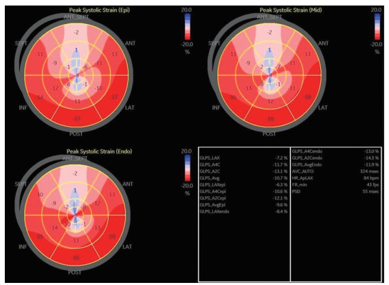
图 1 心内膜分层(外层、中层、内层)纵向应变牛眼示意图
注:负向应变代表收缩期心肌缩短,负向应变越大,颜色标记越接近深红色,颜色越红,表示达峰时间越长。
Figure 1. Schematic diagram of longitudinal strain bovine eye in endocardial stratification (outer, middle, inner)

图 2 GLS-endo、GCS-endo单独及联合预测PCI术后短期预后的ROC曲线
Figure 2. ROC curves of GLS-endo and GCS-endo alone and in combination to predict short-term prognosis after PCI
表 1 对照组与MI组心肌力学参数水平比较(x±s)
Table 1. Comparison of myocardial mechanics parameters between the control group and the MI group (x±s)
组别 例数 LVEF(%) LVEDV(mL) LVESV(mL) GLS-endo GLS-mid GLS-epi GCS-endo GCS-mid GCS-epi 对照组 40 54.19±4.21 108.86±15.06 66.79±7.95 -22.95±1.25 -20.66±1.42 -18.10±1.34 -25.71±1.66 -20.83±1.82 -17.05±1.26 MI组 180 38.95±4.63 116.20±13.95 74.83±12.34 -15.19±3.14 -11.49±2.69 -10.21±2.86 -16.82±3.05 -12.95±2.89 -9.77±2.21 t值 19.112 3.148 3.939 15.340 20.897 17.015 17.835 16.515 20.097 P值 <0.001 0.002 <0.001 <0.001 <0.001 <0.001 <0.001 <0.001 <0.001  下载: 导出CSV
表 2 预后良好组及预后不良组MI患者心肌力学参数水平比较(x±s)
Table 2. Comparison of myocardial mechanical parameters in MI patients with good and poor prognosis (x±s)
组别 例数 LVEF(%) LVEDV(mL) LVESV(mL) GLS-endo GLS-mid GLS-epi GCS-endo GCS-mid GCS-epi 预后良好组 133 39.96±4.59 114.76±19.72 73.96±13.27 -15.97±2.31 -12.06±2.73 -10.76±2.93 -17.83±3.19 -13.54±3.02 -10.23±2.38 预后不良组 47 38.59±4.72 120.29±20.33 77.31±11.91 -12.98±2.96 -9.88±2.59 -8.65±2.51 -13.96±2.81 -11.28±2.71 -8.47±2.24 t值 1.723 1.639 1.527 6.278 3.275 4.398 7.366 4.525 4.424 P值 0.087 0.103 0.129 <0.001 0.001 <0.001 <0.001 <0.001 <0.001
下载: 导出CSV
表 3 预后良好组及预后不良组MI患者临床资料比较
Table 3. Comparison of clinical data of MI patients in the good and poor prognosis groups
项目 预后良好组(n=133) 预后不良组(n=47) 统计量 P值 性别[例(%)] 0.402a 0.526 男性 75(56.39) 29(61.70) 女性 58(43.61) 18(38.30) 年龄(x±s,岁) 55.86±6.15 54.29±6.47 1.484b 0.140 BMI(x±s) 23.05±1.06 23.21±1.13 0.874b 0.384 Killip分级[例(%)] 7.313a 0.007 Ⅰ~Ⅱ级 99(74.44) 25(53.19) Ⅲ级 34(25.56) 22(46.81) 吸烟史[例(%)] 41(30.83) 17(36.17) 0.454a 0.500 饮酒史[例(%)] 46(34.59) 19(40.43) 0.513a 0.474 脑卒中史[例(%)] 13(9.77) 8(17.02) 1.770a 0.183 合并症[例(%)] 糖尿病 29(21.80) 14(29.79) 1.217a 0.270 高血压 35(26.32) 17(36.17) 1.642a 0.200 高脂血症 35(26.32) 16(34.04) 1.021a 0.312 病变支数[例(%)] 4.003c <0.001 单只 65(48.87) 9(19.15) 双支 41(30.83) 16(34.04) 三支及以上 27(20.30) 22(46.81) 心肌梗死面积[例(%)] 2.218c 0.330 小面积梗死 43(32.33) 17(36.17) 中面积梗死 69(51.88) 19(40.43) 大面积梗死 21(15.79) 11(23.40) 开通罪犯血管的时间[例(%)] 9.653a 0.002 <6 h 98(73.68) 23(48.94) 6~12 h 35(26.32) 24(51.06) 合并术前用药[例(%)] 他汀类 106(79.70) 35(74.47) 0.560a 0.454 双抗 87(65.41) 29(61.70) 0.209a 0.648 替罗非班 119(89.47) 40(85.11) 0.643a 0.423 肌酸激酶(x±s,U/L) 1 416.65±306.96 1 495.6±335.45 1.479b 0.141 肌酸激酶同工酶(x±s,U/L) 288.87±40.33 296.79±39.56 1.163b 0.246 脑利尿钠肽(x±s,ng/L) 336.59±53.29 577.54±95.33 3.442b 0.001 靶血管长度(x±s,mm) 2.85±0.49 2.94±0.54 1.054b 0.294 靶血管直径(x±s,mm) 23.95±4.36 24.61±4.57 0.881b 0.380 植入支架数量(x±s,个) 1.65±0.24 1.82±0.21 2.698b 0.008 心率(x±s,次/min) 75.95±11.26 77.16±10.79 0.640b 0.523 收缩压(x±s,mmHg) 116.71±14.26 119.26±13.05 1.077b 0.283 舒张压(x±s,mmHg) 75.49±8.16 77.50±8.79 1.422b 0.157 白细胞计数(x±s,×109/L) 7.85±1.86 8.09±1.93 0.753b 0.452 血小板计数(x±s,×109/L) 219.26±41.55 228.54±49.31 1.252b 0.212 总胆固醇(x±s,mmol/L) 4.36±1.09 4.51±1.26 0.778b 0.438 三酰甘油(x±s,mmol/L) 3.89±1.15 3.94±1.07 0.261b 0.795 高密度脂蛋白胆固醇(x±s,mmol/L) 1.04±0.33 1.07±0.26 0.564b 0.573 低密度脂蛋白胆固醇(x±s,mmol/L) 2.79±0.87 2.93±0.75 0.981b 0.328 天门冬氨酸氨基转移酶(x±s,U/L) 116.24±38.59 121.73±33.45 0.867b 0.387 丙氨酸氨基转移酶(x±s,U/L) 50.49±17.26 53.21±15.76 0.948b 0.344 注:a为χ2值,b为t值, c为Z值。1 mmHg=0.133 kPa。
下载: 导出CSV
表 4 MI患者PCI术后短期预后影响因素的多因素logistic回归分析
Table 4. Multivariate logistic regression analysis affecting the short-term prognosis of MI patients after PCI
变量 B SE Waldχ2 P值 OR值 95% CI Killip分级 0.227 0.095 5.710 <0.001 1.255 1.042~1.512 病变支数 0.605 0.325 3.465 0.091 1.831 0.969~3.463 开通罪犯血管的时间 0.596 0.245 5.918 <0.001 1.815 1.123~2.934 脑利尿钠肽 0.596 0.334 3.603 0.083 1.815 0.981~3.358 植入支架数量 0.841 0.483 3.032 0.154 2.319 0.900~5.976 GLS-endo -0.409 0.201 4.141 <0.001 0.664 0.448~0.985 GLS-mid -0.565 0.305 3.432 0.076 0.568 0.313~1.033 GLS-epi -0.395 0.221 3.195 0.095 0.674 0.437~1.039 GCS-endo -0.245 0.119 4.239 <0.001 0.783 0.620~0.988 GCS-mid -0.664 0.340 3.814 0.115 0.515 0.264~1.002 GCS-epi -0.492 0.254 3.752 0.073 0.611 0.372~1.006 注:变量赋值如下,Killip分级,Ⅲ级=1,Ⅰ~Ⅱ级=0;病变支数,三支及以上=2,双支=1,单支=0;开通罪犯血管的时间,6~12 h=1,<6 h=0;其余连续变量均以实际值赋值。
下载: 导出CSV
表 5 心肌力学参数对PCI术后短期预后的预测价值
Table 5. The predictive value of myocardial mechanical parameters for short-term prognosis after PCI
指标 截断值 灵敏度
(%)特异度
(%)AUC 95% CI 约登指数 GLS-endo 13.41 77.78 67.41 0.767 0.699~0.827 0.452 GCS-endo 14.73 60.00 83.70 0.714 0.642~0.779 0.437 联合 1.76a 91.11 84.44 0.870 0.812~0.915 0.756 注:a联合预测采用LogP模式进行拟合;联合诊断最佳截断值依据Log(P/1-P)模型生成。
下载: 导出CSV
-
[1] SAGRIS M, ANTONOPOULOS A S, THEOFILIS P, et al. Risk factors profile of young and older patients with myocardial infarction[J]. Cardiovasc Res, 2022, 118(10): 2281-2292. doi: 10.1093/cvr/cvab264 [2] SAITO Y, OYAMA K, TSUJITA K, et al. Treatment strategies of acute myocardial infarction: updates on revascularization, pharmacological therapy, and beyond[J]. J Cardiol, 2023, 81(2): 168-178. doi: 10.1016/j.jjcc.2022.07.003 [3] FRAMPTON J, ORTENGREN A R, ZEITLER E P. Arrhythmias after acute myocardial infarction[J]. Yale J Biol Med, 2023, 96(1): 83-94. doi: 10.59249/LSWK8578 [4] 丁平, 楚曌涵, 郭晓峰, 等. 正念减压式抗阻训练对急性心肌梗死PCI术后患者心功能及冠脉微循环的影响研究[J]. 医药论坛杂志, 2024, 45(13): 1434-1438.DING P, CHU Z H, GUO X F, et al. Effect of mindfulness-based stress reduction resistance training on cardiac function and coronary microcirculation in patients with acute myocardial infarction after PCI[J]. Journal of Medical Forum, 2024, 45(13): 1434-1438. [5] 李慧君, 李思, 白旭鹏, 等. 沙库巴曲缬沙坦对急性ST段抬高型心肌梗死PCI术后心功能影响的临床观察[J]. 北京医学, 2023, 45(3): 193-197.LI H J, LI S, BAI X P, et al. Clinical Observation on the effect of sacubitril/valsartan on cardiac function after PCI in acute ST-elevation myocardial infarction[J]. Journal of New Medicine, 2023, 45(3): 193-197. [6] YUDA S. Current clinical applications of speckle tracking echocardiography for assessment of left atrial function[J]. J Echocardiogr, 2021, 19(3): 129-140. doi: 10.1007/s12574-021-00519-8 [7] MORARIU V I, ARNAUTU D A, MORARIU S I, et al. 2D speckle tracking: a diagnostic and prognostic tool of paramount importance[J]. Eur Rev Med Pharmacol Sci, 2022, 26(11): 3903-3910. [8] MANDOLI G E, BORRELLI C, CAMELI M, et al. Speckle tracking echocardiography in heart failure development and progression in patients with apneas[J]. Heart Fail Rev, 2022, 27(5): 1869-1881. doi: 10.1007/s10741-021-10197-4 [9] 中华医学会心血管病学分会, 中华心血管病杂志编辑委员会. 急性ST段抬高型心肌梗死诊断和治疗指南[J]. 中华心血管病杂志, 2015, 43(5): 380-393.Chinese Society of Cardiology, Editorial Board of Chinese Journal of Cardiology. Guideline for diagnosis and treatment of patients with ST-elevation myocardial infarction[J]. Chinese Journal of Cardiology, 2015, 43(5): 380-393. [10] 中华医学会心血管病学分会介入心脏病学组, 中国医师协会心血管内科医师分会血栓防治专业委员会, 中华心血管病杂志编辑委员会. 中国经皮冠状动脉介入治疗指南(2016)[J]. 中华心血管病杂志, 2016, 44(5): 382-400.Interventional Cardiology Group, Chinese Society of Cardiology, Chinese Medical Association Branch of Cardiovascular Physicians, Thrombus Prevention and Treatment Committee, Chinese Journal of Cardiology Editorial Committee. Chinese Guidelines for percutaneous coronary intervention (2016)[J]. Chinese Journal of Cardiology, 2016, 44(5): 382-400. [11] 董扬, 张芬, 王文标, 等. 老年急性心肌梗死患者PCI和非PCI治疗预后比较[J]. 中华全科医学, 2017, 15(4): 721-723. doi: 10.16766/j.cnki.issn.1674-4152.2017.04.053DONG Y, ZHANG F, WANG W B, et al. Comparison of prognosis between PCI and non-PCI treatment in elderly patients with acute myocardial infarction[J]. Chinese Journal of General Practice, 2022, 15(4): 721-723. doi: 10.16766/j.cnki.issn.1674-4152.2017.04.053 [12] NISHIHIRA K, KURIYAMA N, KADOOKA K, et al. Outcomes of elderly patients with acute myocardial infarction and heart failure who undergo percutaneous coronary intervention[J]. Circ Rep, 2022, 4(10): 474-481. doi: 10.1253/circrep.CR-22-0048 [13] 王志伟, 王真真, 刘畅, 等. 排除国际标准化比值的终末期肝病模型评分对急性心肌梗死后室间隔穿孔患者行经皮介入封堵术后预后的预测价值[J]. 中华实用诊断与治疗杂志, 2023, 37(2): 156-161.WANG Z W, WANG Z Z, LIU C, et al. Value of model for end-stage liver disease excluding international normalized ratio to the prediction of prognosis of post-myocardial infarction ventricular septal rupture after percutaneous transcatheter closure[J]. Journal of Chinese Practical Diagnosis and Therapy, 2023, 37(2): 156-161. [14] 邓冰晴, 王开权, 李金国, 等. 三维超声斑点追踪技术定量评估维持性血液透析患者左心室旋转的价值[J]. 中华实用诊断与治疗杂志, 2018, 32(1): 91-93.DENG B Q, WANG K Q, LI J G, et al. Value of three-dimensional ultrasound speckle tracking technique to the quantitative evaluation of left ventricular rotation in patients receiving maintenance hemodialysis[J]. Journal of Chinese Practical Diagnosis and Therapy, 2018, 32(1): 91-93. [15] SADE L E, JOSHI S S, CAMELI M, et al. Current clinical use of speckle-tracking strain imaging: insights from a worldwide survey from the European Association of Cardiovascular Imaging (EACVI)[J]. Eur Heart J Cardiovasc Imaging, 2023, 24(12): 1583-1592. doi: 10.1093/ehjci/jead170 [16] 崔秀秀, 董彧, 王颖, 等. 三维斑点追踪技术对无心肌梗死冠心病三支血管病变患者左心室功能的评价[J]. 中国医师杂志, 2022, 24(5): 739-744.CUI X X, DONG Y, WANG Y, et al. Three-dimensional speckle tracking echocardiography in evaluating left ventricular function in patients with triple vessel coronary artery disease without myocardial infarction[J]. Journal of Chinese Physician, 2022, 24(5): 739-744. [17] 赵瑞环. 超声斑点追踪技术评价急性心梗PCI前后心肌力学改变[D]. 太原: 山西医科大学, 2018.ZHAO R H. Evaluation of myocardial mechanical changes before and after PCI by ultrasonic speckle tracking technique[D]. Taiyuan: Shanxi Medical University, 2018. [18] 米佳, 王佳强, 闫娟. 二维斑点追踪超声心动图在评价冠脉多支病变患者左心功能中的作用[J]. 中国医师杂志, 2019, 21(3): 401-405, 409.MI J, WANG J Q, YAN J. The role of two-dimensional speckle tracking echocardiography in evaluating left ventricular function of patients with multi-vessel coronary artery disease[J]. Journal of Chinese Physician, 2019, 21(3): 401-405, 409. [19] GAO Q, LIU P, LV T T, et al. Utility of speckle-tracking echocardiography for predicting atrial fibrillation following ischemic stroke: a systematic review and meta-analysis[J]. Int J Cardiovasc Imaging, 2022, 38(8): 1771-1780. doi: 10.1007/s10554-022-02570-7 [20] ZHOU F O, YUAN H, SUN J D, et al. Two-dimensional speckle tracking imaging cardiac motion-based quantitative evaluation of global longitudinal strain among patients with coronary heart disease and functions of left ventricular ischemic myocardial segment[J]. Int J Cardiovasc Imaging, 2024, 40(2): 351-359. [21] HATORI M, SAKAKURA K, TANIGUCHI Y, et al. Factors associated with in-hospital death in patients with Killip class 3 acute myocardial infarction[J]. Int Heart J, 2021, 62(4): 756-763. doi: 10.1536/ihj.21-078 -


 点击查看大图
计量
- 文章访问数: 11
- HTML全文浏览量: 2
- PDF下载量: 0
- 被引次数: 0
