

Correlation between MRI imaging characteristics of non-mass-like breast cancer and Ki-67 expression levels in non-mass breast cancer
-
摘要:
目的 探讨非肿块型乳腺癌MRI特征与Ki-67表达相关性,为术前无创评估肿瘤增殖活性提供依据。 方法 收集2020年1月—2023年12月在安徽医科大学第二附属医院就诊并经病理确诊的51例非肿块型乳腺癌患者的资料,根据术后病理分为Ki-67低表达组(15例)和Ki-67高表达组(36例),比较2组临床及MRI特征[平均表观扩散系数(ADC)值、腋窝淋巴结转移、强化分布、内部强化方式、时间-信号强度曲线(TIC)]。 结果 2组患者年龄、平均ADC值、强化分布、内部强化间差异均无统计学意义(P>0.05),内部强化以簇环状强化为主(47.06%,24/51)。高表达组腋窝淋巴结转移率显著高于低表达组[58.33%(21/36)vs. 26.67%(4/15), P=0.041]。高表达组TIC曲线Ⅰ型1例、Ⅱ型15例、Ⅲ型20例,低表达组TIC曲线Ⅰ型3例、Ⅱ型9例、Ⅲ型3例,2组曲线分布差异有统计学意义(P=0.018),其中TIC Ⅰ型和Ⅲ型曲线间Ki-67表达差异有统计学意义(经Bonferroni校正, P < 0.017)。腋窝淋巴结转移率与Ki-67表达呈正相关关系(Kendall,S tau-b=0.289,P=0.041),病灶平均ADC值与其呈负相关关系(r=-0.334,P=0.017),TIC曲线类型与其呈正相关关系(r=0.303,P=0.031)。腋窝淋巴结转移及TIC曲线预测Ki-67表达水平的AUC分别为0.658、0.678,两者联合预测的AUC为0.761(灵敏度为0.861,特异度为0.667)。 结论 非肿块型乳腺癌以簇环状强化多见,腋窝淋巴结转移及TIC曲线对Ki-67表达具有一定的预测效能,联合指标可提升预测效能,为术前评估肿瘤增殖活性提供参考。 Abstract:Objective To investigate the correlation between MRI features of non-mass breast cancer and Ki-67 expression, providing a basis for non-invasive preoperative assessment of tumor proliferative activity. Methods From January 2020 to December 2023, fifty-one patients with pathologically confirmed non-mass breast cancer were enrolled at the Second Affiliated Hospital of Anhui Medical University. Based on postoperative pathology, they were stratified into a low Ki-67 expression group (n=15) and a high-expression group (n=36). Comparisons included clinical data and MRI features [mean apparent diffusion coefficient (ADC) value, axillary lymph node metastasis, enhancement distribution, internal enhancement pattern, time-intensity curve (TIC) curve]. Results No significant differences were observed in age, mean ADC value, enhancement distribution, or internal enhancement between groups (P>0.05), with clustered ring enhancement predominating (47.06%, 24/51). The high-expression group showed significantly higher axillary lymph node metastasis rates than the low-expression group [58.33%(21/36) vs. 26.67% (4/15), P=0.041].TIC curves in the high Ki-67 group were categorized as type Ⅰ (1 case), type Ⅱ(15 cases), and type Ⅲ (20 cases), while the low Ki-67 group showed type Ⅰ (3 cases), type Ⅱ (9 cases), and type Ⅲ (3 cases). TIC distribution differed significantly between groups (P=0.018), with statistically significant differences in Ki-67 expression between type Ⅰ and Ⅲ curves (Bonferroni-corrected, P < 0.017). Axillary lymph node metastasis rate positively correlated with Ki-67 expression (Kendall' s tau-b=0.289, P=0.041). mean ADC value negatively correlated (r=-0.334, P=0.017), and TIC curve type positively correlated (r=0.303, P=0.031). The combined AUC of axillary lymph node metastasis and TIC curves for predicting Ki-67 expression was 0.761 (sensitivity 0.861, specificity 0.667), outperforming individual indicators (AUC: 0.658, 0.678). Conclusion Non-mass breast cancer frequently exhibits clustered ring enhancement. Axillary lymph node metastasis and TIC curves correlate with Ki-67 expression, and their combination improves predictive efficacy, offering value for preoperative assessment of tumor proliferative activity. -
Key words:
- Non-mass breast cancer /
- ADC values /
- TIC curve type /
- Ki-67 /
- Magnetic resonance imaging
-

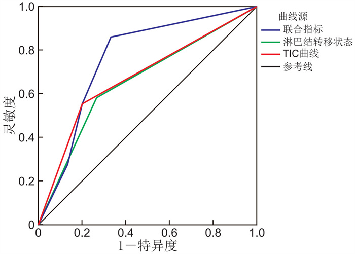
图 1 不同指标预测非肿块型乳腺癌患者Ki-67高表达的ROC曲线
Figure 1. ROC curves of different indicators predicting high Ki-67 expression in patients with non-mass breast cancer

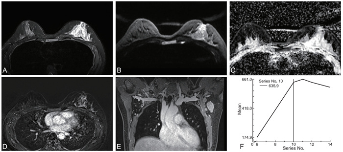
图 2 非肿块型乳腺癌患者MRI影像(Ki-67高表达,90%)
注:患者,女性,40岁,A为T2WI序列,左乳病灶呈高信号;B为DWI序列,病灶呈稍高信号;C为ADC图,病灶呈稍低信号,ADC值约1.234×10⁻³mm2/s;D为MRI增强横断面,左乳病灶呈散在分布集簇状强化灶;E为MRI增强冠状位,腋窝淋巴结转移;F为TIC曲线,Ⅲ型。
Figure 2. MRI images of a patient with non-mass breast cancer showing high Ki-67 expression (90%)

图 3 非肿块型乳腺癌患者MRI影像(Ki-67低表达, 15%)
注:患者,女性,50岁,A为T2WI序列,左乳病灶呈高信号;B为DWI序列,病灶呈稍高信号;C为ADC图,病灶呈稍低信号,ADC值约1.035×10⁻³mm2/s;D为MRI增强横断面,左乳病灶呈段样分布簇环状强化灶;E为MRI增强冠状位,无腋窝淋巴结转移;F为TIC曲线,Ⅱ型。
Figure 3. MRI images of a patient with non-mass breast cancer showing low Ki-67 expression (15%)
表 1 2组非肿块型乳腺癌患者临床及MRI相关资料比较
Table 1. Comparison of clinical and MRI data between two groups of patients with non-mass breast cancer
项目 Ki-67低表达组(n=15) Ki-67高表达组(n=36) 统计量 P值 年龄(x±s,岁) 45.87±7.21 45.19±8.92 0.258a 0.798 肿瘤长径(x±s, cm) 4.21±1.74 5.25±2.20 1.639a 0.108 ADC值(x±s,10-3mm2/s) 1.061±0.113 0.998±0.128 1.673a 0.101 腋窝淋巴结转移[例(%)] 4(26.67) 21(58.33) 4.165b 0.041 强化分布方式[例(%)] 4.493b 0.310 局灶强化 1(6.67) 3(8.33) 段样强化 1(6.67) 3(8.33) 区域强化 8(53.33) 9(25.00) 多区域强化 4(26.67) 19(52.78) 弥漫分布 1(6.67) 2(5.56) 内部强化方式[例(%)] 5.846b 0.102 均匀 1(6.67) 3(8.33) 不均匀 8(53.33) 7(19.44) 簇环状 4(26.67) 20(55.56) 集簇状 2(13.33) 6(16.67) TIC曲线类型[例(%)] 7.383b 0.018 Ⅰ型 3(20.00) 1(2.78) Ⅱ型 9(60.00) 15(41.67) Ⅲ型 3(20.00) 20(55.56) 注:a为t值,b为χ2值。  下载: 导出CSV
表 2 MRI参数对非肿块型乳腺癌Ki-67高表达的预测价值
Table 2. Predictive value of MRI parameters for high Ki-67 expression in non-mass breast lesions
参数 AUC 95% CI P值 灵敏度 特异度 腋窝淋巴结转移状态 0.658 0.495~0.821 0.038 0.583 0.733 TIC类型 0.678 0.520~0.835 0.017 0.556 0.800 腋窝淋巴结转移状态+TIC类型 0.761 0.600~0.922 0.004 0.861 0.667
下载: 导出CSV
-
[1] HAN B F, ZHENG R S, ZENG H M, et al. Cancer incidence and mortality in China, 2022[J]. J Natl Cancer Cent, 2024, 4(1): 47-53. [2] PARK K W, PARK S, SHON I, et al. Non-mass lesions detected by breast US: stratification of cancer risk for clinical management[J]. Eur Radiol, 2021, 31(3): 1693-1706. doi: 10.1007/s00330-020-07168-y [3] 于鹏丽, 孔文韬, 薛海燕, 等. 声触诊组织成像量化技术鉴别诊断非肿块型乳腺病变良恶性的临床价值[J]. 肿瘤影像学, 2022, 31(6): 575-580.YU P L, KONG W T, XUE H Y, et al. The clinical value of virtual touch tissue imaging quantification technology in differential diagnosis of benign and malignant non-mass breast lesions[J]. Oncoradiology, 2022, 31(6): 575-580. [4] 宋旗, 胡友庭, 杨泳, 等. 乳腺癌组织细胞核增殖抗原的异常表达与病理特征相关性[J]. 中华实验外科杂志, 2020, 37(2): 317-320.SONG Q, HU Y T, YANG Y, et al. Correlation between expression of proliferation cell nuclear antigen protein and pathological characteristics in breast cancer[J]. Chinese Journal of Experimental Surgery, 2020, 37(2): 317-320. [5] 陈佳佳, 孟利伟, 李星云, 等. 乳腺癌超声特征和ER、PR、CerbB-2、Ki-67阳性表达的相关性研究[J]. 中华全科医学, 2021, 19(10): 1721-1724. doi: 10.16766/j.cnki.issn.1674-4152.002141CHEN J J, MENG L W, LI X Y, et al. Relationship between ultrasound features and expression of ER, PR, CerbB-2 and Ki-67 in breast cancer[J]. Chinese Journal of General Practice, 2021, 19(10): 1721-1724. doi: 10.16766/j.cnki.issn.1674-4152.002141 [6] ZHANG L Y, HAO J S, GUO J, et al. Predicting of Ki-67 expression level using diffusion-weighted and synthetic magnetic resonance imaging in invasive ductal breast cancer[J]. Breast J, 2023, 2023: 6746326. DOI: 10.1155/2023/6746326. [7] 中国抗癌协会乳腺癌专业委员会, 中华医学会肿瘤学分会乳腺肿瘤学组. 中国抗癌协会乳腺癌诊治指南与规范(2024年版)[J]. 中国癌症杂志, 2023, 33(12): 1092-1187.The society of breast cancer China Anti-Cancer association, Breast oncology group of the oncology branch of the Chinese Medical Association. Guidelines for breast cancer diagnosis and treatment by China Anti-cancer Association(2024 edition)[J]. China Oncology, 2023, 33(12): 1092-1187. [8] 李阳, 李玉梅, 邓军. 等. 乳腺癌新辅助化疗前后ER、PR、Her-2和Ki-67的变化与化疗疗效的关系分析[J]. 中华全科医学, 2024, 22(9): 1500-1503. doi: 10.16766/j.cnki.issn.1674-4152.003668LI Y, LI Y M, DENG J, et al. Relationship between the changes of ER, PR, Her-2 and Ki-67 before and after neoadjuvant chemotherapy and chemotherapy efficacy in breast cancerr[J]. Chinese Journal of General Practice, 2024, 22(9): 1500-1503. doi: 10.16766/j.cnki.issn.1674-4152.003668 [9] 陈艳虹, 王丽君, 罗冉, 等. 乳腺癌腋窝淋巴结转移与原发灶MRI及临床病理特征的相关性研究[J]. 中国医学计算机成像杂志, 2022, 28(5): 484-490.CHEN Y H, WANG L J, LUO R, et al. Investigation on the correlation between axillary lymph node metastasis and preoperative MRI and clinicopathological features of invasive breast Cancer[J]. China Computer Medical Imaging, 2022, 28(5): 484-490. [10] 李琳, 崔文静, 崔延安, 等. 动态增强MRI在乳腺非肿块样强化良恶性病变鉴别诊断中的临床应用研究[J]. 东南大学学报(医学版), 2023, 42(6): 903-908.LI L, CUI W J, CUI Y A, et al. Clinical application of DCE-MRI in differential diagnosis of benign and malignant breast lesions with non-mass enhancement[J]. Journal of Southeast University(medical Science Edition), 2023, 42(6): 903-908. [11] 周鑫, 王珍, 付雨菲. 乳腺非肿块样强化良恶性病变DCE-MRI表现及其诊断价值分析[J]. 中国CT和MRI杂志, 2024, 22(7): 118-120.ZHOU X, WANG Z, REN Y F. Analysis of DCE-MRI features and fiagnostic value of non-mass-enhancing benign and malignant breast lesions[J]. Chinese Jouranl of CT and MRI, 2024, 22(7): 118-120. [12] 崔晓琳, 杨正汉, 张洁, 等. 乳腺簇状环形强化非肿块病变的MRI表现与病理学对照研究[J]. 医学影像学杂志, 2019, 29(11): 1900-1904.CUI X L, YANG Z H, ZHANG J, et al. Breast lesions presented as cluster ring enhancement on MRI: comparison of imaging features and pathology[J]. Journal of Medical Imaging, 2019, 29(11): 1900-1904. [13] 刘靓, 朱丹, 沈晶, 等. 多模态MRI技术在乳腺非肿块性强化病变良恶性鉴别中的临床研究[J]. 中国临床医学影像杂志, 2020, 31(1): 15-19.LIU L, ZHU D, SHEN J, et al. Clinical study of multi-mode MRI in differentiating benign and malignant breast non-mass-like enhancement lesions[J]. Journal of China Clinic Medical Imaging, 2020, 31(1): 15-19. [14] 黄瑞岁, 丁可, 林彬, 等. 多模态MRI对非肿块型乳腺癌的诊断价值研究[J]. 医学影像学杂志, 2021, 31(6): 997-1001.HUANG R S, DING K, LIN B, et al. Study on the diagnostic value of multi-modal MRI in non-mass breast cancer[J]. Journal of Medical Imaging, 2021, 31(6): 997-1001. [15] 廖建勇, 杜静波, 刘迎新, 等. 基于动态增强磁共振成像和扩散加权成像预测乳腺癌Ki-67表达水平的可行性研究[J]. 中国CT和MRI杂志, 2023, 21(9): 94-96.LIAO J Y, DU J B, LIU Y X, et al. Feasibility of predicting Ki-67 expression level in breast cancer based on dynamic contrast-enhanced magnetic resonance imaging and diffusion-weighted imaging[J]. Chinese Jouranl of CT and MRI, 2023, 21(9): 94-96. [16] 姚明, 程流泉, 李梦露, 等. 浸润性乳腺癌肿块与非肿块强化方式的表观扩散系数分布特点[J]. 中国医学影像学杂志, 2020, 28(2): 90-94.YAO M, CHENG L Q, LI M L, et al. Distribution characteristics of apparent diffusion coefficient difference between mass and non mass-enhancement of Invasive breast bancer[J]. Chinese Journal of Medical Imaging, 2020, 28(2): 90-94. [17] 吴向东, 田林, 郝晓鹏. 肿块强化与非肿块强化乳腺癌患者的临床和病理特征对比分析[J]. 中国临床新医学, 2022, 15(11): 1003-1007.WU X D, TIAN L, HAO X P. Analysis on the clinical and pathological features the patients with mass enhanced breast cancer and those with non-mass enhanced breast bancer[J]. Chinese Journal of New Clinical Medicine, 2022, 15(11): 1003-1007. [18] AVENDANO D, MARINO M A, LEITHNER D, et al. Limited role of DWI with apparent diffusion coefficient mapping in breast lesions presenting as non-mass enhancement on dynamic contrast-enhanced MRI[J]. Breast Cancer Res, 2019, 21(1): 136. DOI: 10.1186/s13058-019-1208-y. -


 点击查看大图
计量
- 文章访问数: 4
- HTML全文浏览量: 1
- PDF下载量: 0
- 被引次数: 0
