

Congenital portosystemic shunts: 2 cases report and review of literature
-
摘要: 增加临床医生对先天性门体分流(CPSS)的认识,重视患儿病史细节,达到尽早发现、制定个性化治疗方案的目的。回顾分析近10年在安徽省儿童医院就诊并治疗的2例新生儿CPSS临床资料,并以“先天性门体分流”“先天性门静脉分流”“新生儿”“Congenital portosystemic shunts”“Abernethy syndrome”“Infants”“Case report”为关键词检索2010年1月1日—2022年12月31日发表在中国知网、万方数据库、维普网数据库、PubMed、GeenMedical、Web of Science的中英文文献,通过去重、删除资料不全文献,共纳入相关文献10篇,共118例患者,进行文献复习。分析其临床表现、相关合并症、治疗及预后,以增强临床医师对先天性门静脉分流的认识,提高对该病的诊断和管理能力。
-
关键词:
- 先天性门体分流 /
- 新生儿 /
- Abernethy畸形 /
- 自发闭合 /
- 文献复习
Abstract: To increase clinicians' awareness of congenital portosystemic shunts, pay attention to the details of the child's medical history, achieve early detection and develop individualized treatment plans. Retrospective analysis of clinical data of 2 cases of CPSS in children seen and treated at Anhui Children's Hospital in the past 10 years. Then we systematically searched relevant studies in Chinese and English literature between January 1, 2010 and December 31, 2022 in databases of China Knowledge Network, Wanfang database, Vipshop database, PubMed, GeenMedical, Web of Science using congenital portosystemic shunts, Abernethy syndrome, infants, case report as the keywords.After removing the duplicates and those with incomplete data, we conducted a review of 10 studies with 118 cases. Our analysis of the clinical manifestations, diagnosis and treatment of these cases may help to improve clinicians' understanding of congenital portosystemic shunts and their ability to diagnose and manage the condition.-
Key words:
- Congenital portosystemic shunts /
- Infants /
- Abernethy syndrome /
- Spontaneous closure /
- Literature review
-

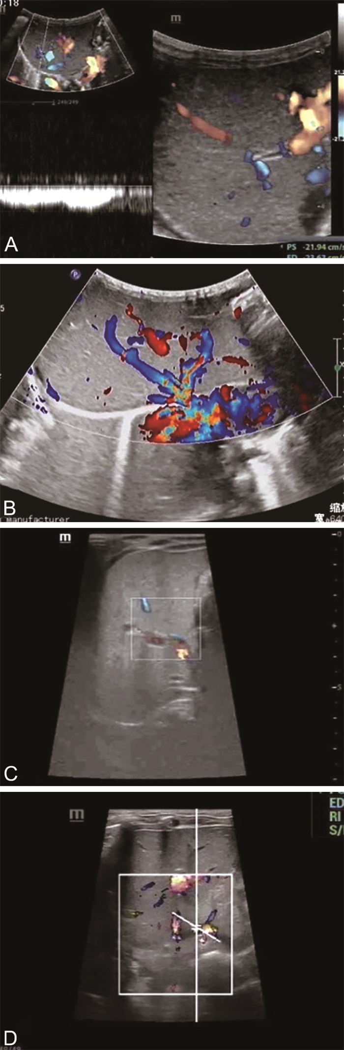
图 1 2例新生儿先天性门体分流患儿肝胆胰脾超声检查结果
注:A为病例1患儿21 d检查结果,肝静脉流速约23 cm/s,肝右叶似扫及一处门静脉血流流入肝静脉,此处肝静脉流速显著增快,约43 cm/s;B为病例1患儿1月龄复查结果,肝脏肋下约1.7 cm,门静脉与肝静脉均未见明显扩张,流速正常;C为病例1患儿2月龄复查结果,门静脉与肝静脉均未见明显扩张,肝内血管走形清晰,异常分流已消失;D为病例2出生后复查结果,门静脉流速偏低,肝中静脉流速增快,脐静脉已闭合。
Figure 1. Ultrasound examination results of liver, biliary, pancreas, and spleen in 2 newborn children with congenital portal shunt
表 1 新生儿门静脉分流病例临床诊断结果(例)
Table 1. Clinical diagnosis results of neonatal cases with ductus venosus shunt (cases)
第一作者 发表年份 例数 肝内/肝外分流(例) 诊断年龄(范围) 产前诊断例数(例) 季敏[2] 2017 8 0/8 (4.17±3.64)岁(3个月~10岁) 0 向永华[3] 2020 28 24/4 肝内分流(1.1±0.2)个月 0 肝外分流3.99岁(1个月~7.90岁) 肖云彬[4] 2019 5 3/2 肝内分流1.67个月(1~3个月) 0 肝外分流9个月,4.4岁 0 谢恩萍[5] 2017 1 1/0 4 d 0 XU S H[6] 2021 16 13/3 16 d(产前24周~12个月) 3 DIPAOLA F[7] 2020 11 3/8 10岁(1个月~26岁) 0 GONG Y[8] 2015 11 4/7 4个月(5 d~10岁) 0 KIM M J[9] 2012 10 9/1 1.03岁(8 d~8岁) 4 PECˇEK J[10] 2020 5 2/3 22个月(小于1个月~14岁) 0 BLANC T[11] 2014 23 0/23 10岁(2个月~17岁) 8 合计 118 59/59 15  下载: 导出CSV
表 2 新生儿先天性门静脉分流病例临床表现及合并症(n=120)
Table 2. Clinical manifestations and complications of congenital portal vein shunt in newborns(n=120)
项目 类别 例(%) 临床表现 高氨血症 23(19.17) 消化道出血 19(15.83) 肺动脉高压 17(14.17) 肝性脑病 15(12.50) 血小板减少 12(10.00) 合并症 先天性心脏病 51(42.50) 肝脏病变 41(34.17) 精神异常/智力低下 12(10.00) 血管瘤 6(5.00) 染色体异常 5(4.17) 骨代谢异常 4(3.33) 脏器异位症 3(2.50)
下载: 导出CSV
-
[1] WANG Y, YAN Y N, YANG Z J, et al. Prenatal diagnosis of congenital portosystemic shunt: a single-center study[J]. J Obstet Gynaecol Res, 2020, 46(10): 1988-1993. doi: 10.1111/jog.14403 [2] 季敏, 吴先胜, 龚英, 等. 并发消化道出血的先天性门体分流畸形8例影像学特征病例系列报告[J]. 中国循证儿科杂志, 2017, 12(5): 357-361.JI M, WU X S, GONG Y, et al. Imaging features of 8 cases of congenital portosystemic shunt with gastrointestinal hemorrhage: Case series report[J]. Chinese Journal of Evidence-Based Pediatrics, 2017, 12(5): 357-361. [3] 向永华, 金科, 徐和平, 等. 儿童先天性门体静脉分流临床及CT表现[J]. 放射学实践, 2020, 35(10): 1320-1323.XIANG Y H, JIN K, XU H P, et al. Clinical and CT findings of congenital portosystemic venous shunts in children[J]. Radiologic Practice, 2020, 35(10): 1320-1323. [4] 肖云彬, 曾云红, 肖政辉, 等. 先天性门体静脉分流相关性肺动脉高压5例临床分析[J]. 临床儿科杂志, 2019, 37(12): 946-949.XIAO Y B, ZENG Y H, XIAO Z H, et al. Clinical analysis of pulmonary arterial hypertension associated with congenital portosystemic shunt in children[J]. Journal of Clinical Pediatrics, 2019, 37(12): 946-949. [5] 谢恩萍, 张国庆, 步军. 新生儿先天性门体静脉分流合并髂动脉-脐静脉瘘一例报告并文献复习[J]. 中华新生儿科杂志(中英文), 2017, 32(4): 287-290.XIE E P, ZHANG G Q, BU J, et al. Neonatal congenital portosystemic shunt complicated with iliac artery-umbilical vein fistula: a case report and literature review[J]. Chinese Journal of Neonatology, 2017, 32(4): 287-290. [6] XU S H, ZHANG P, HU L Y, et al. Case Report: clinical features of congenital portosystemic shunts in the neonatal period[J]. Front Pediatr, 2021, 9: 778791. DOI: 10.3389/fped.2021.778791. [7] DIPAOLA F, TROUT A T, WALTHER A E, et al. Congenital portosystemic shunts in children: associations, complications, and outcomes[J]. Dig Dis Sci, 2020, 65(4): 1239-1251. doi: 10.1007/s10620-019-05834-w [8] GONG Y, ZHU H, CHEN J, et al. Congenital portosystemic shunts with and without gastrointestinal bleeding-case series[J]. Pediatr Radiol, 2015, 45(13): 1964-1971. doi: 10.1007/s00247-015-3417-6 [9] KIM M J, KO J S, SEO J K, et al. Clinical features of congenital portosystemic shunt in children[J]. Eur J Pediatr, 2012, 171(2): 395-400. doi: 10.1007/s00431-011-1564-9 [10] PEČEK J, FISTER P, HOMAN M. Abernethy syndrome in Slovenian children: five case reports and review of literature[J]. World J Gastroenterol, 2020, 26(37): 5731-5744. doi: 10.3748/wjg.v26.i37.5731 [11] BLANC T, GUERIN F, FRANCHI-ABELLA S, et al. Congenital portosystemic shunts in children: a new anatomical classification correlated with surgical strategy[J]. Ann Surg, 2014, 260(1): 188-198. doi: 10.1097/SLA.0000000000000266 [12] FRANCHI-ABELLA S, GONZALES E, ACKERMANN O, et al. Congenital portosystemic shunts: diagnosis and treatment[J]. Abdom Radiol (NY), 2018, 43(8): 2023-2036. doi: 10.1007/s00261-018-1619-8 [13] PAPAMICHAILl M, PIZANIAS M, HEATON N. Congenital portosystemic venous shunt[J]. Eur J Pediatr, 2018, 177(3): 285-294. doi: 10.1007/s00431-017-3058-x [14] SANADA Y, MIZUTA K. Congenital absence of the portal vein: translated version[J]. J Hepatobiliary Pancreat Sci, 2018, 25(8): 359-369. doi: 10.1002/jhbp.572 [15] 陈骊珠, 张墨, 马巍, 等. 蛋白质组学技术在产前诊断生物标志物筛查中的研究进展[J]. 中华全科医学, 2018, 16(3): 461-464. doi: 10.16766/j.cnki.issn.1674-4152.000128CHEN L Z, ZHANG M, MA W, et al. The main advances in the application of proteomics in prenatal diagnosis[J]. Chinese Journal of General Practice, 2018, 16(3): 461-464. doi: 10.16766/j.cnki.issn.1674-4152.000128 [16] PONCE-DORREGO M D, HERNÁNDEZ-CABRERO T, GARZÓN-MOLL G. Endovascular treatment of congenital portosystemic shunt: a single-center prospective study[J]. Pediatr Gastroenterol Hepatol Nutr, 2022, 25(2): 147-162. doi: 10.5223/pghn.2022.25.2.147 [17] XIAO Y B, LI W F, DENG X C, et al. Ligation of patent ductus venosus in a child with pulmonary arterial hypertension and hypersplenism: a case report[J]. Medicine (Baltimore), 2020, 99(34): e21849. DOI: 10.1097/MD.0000000000021849. [18] MCLIN V A, FRANCHI ABELLA S, DEBRAY D, et al. Congenital portosystemic shunts: current diagnosis and management[J]. J Pediatr Gastroenterol Nutr, 2019, 68(5): 615-622. doi: 10.1097/MPG.0000000000002263 [19] 赵璐, 吴琳. 儿童先天性门-体分流的诊断和治疗[J]. 中华小儿外科杂志, 2020, 41(1): 93-96.ZHAO L, WU L. Diagnosis and management of congenital portosystemic shunts in children[J]. Chinese Journal of Pediatric Surgery, 2020, 41(1): 93-96. [20] LAVERDURE N, LALLIER M, DUBOIS J, et al. Congenital absence of the portal vein: define the portosystemic shunt, avoid liver transplantation[J]. Can Liver J, 2021, 4(3): 322-327. doi: 10.3138/canlivj-2020-0011 [21] YAN X Y, ZHANG P P, QI Z H, et al. Prenatal diagnosis of intrahepatic congenital portosystemic shunt[J]. J Pediatr, 2022, 251: 212-214. doi: 10.1016/j.jpeds.2022.08.028 [22] JIMENEZ-GOMEZ J, GÜIZZO J R, BETANCOURTH ALVARENGA J, et al. Correlation of prenatal and postnatal diagnosis in umbilical-portal-systemic venous shunts[J]. Eur J Pediatr Surg, 2023, 33(1): 90-95. doi: 10.1055/s-0042-1760379 -

 点击查看大图
计量
- 文章访问数: 293
- HTML全文浏览量: 231
- PDF下载量: 14
- 被引次数: 0
