

-
-

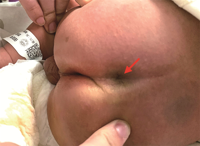
图 1 骶尾部可见凹陷样小孔(红色箭头)
Figure 1. The sacrococcygeal cavity is seen as a pitted hole (red arrow)

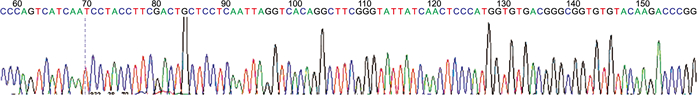
图 2 脑脊液高通量测序技术结果(大芬戈尔德菌)
Figure 2. Results of high-throughput sequencing of cerebrospinal fluid (Fingolderia magnum)

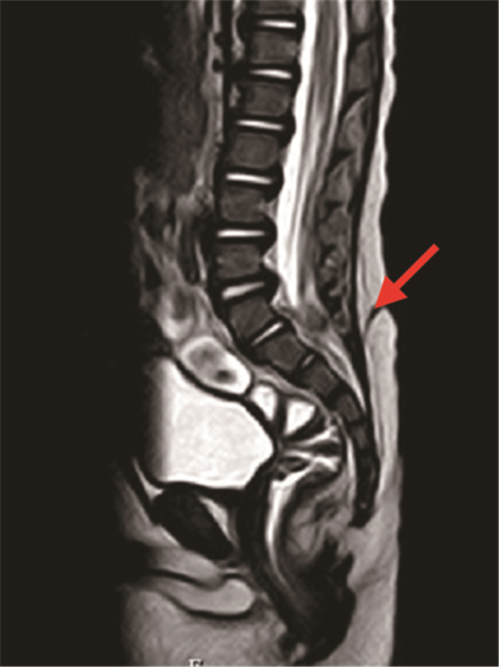
图 3 腰骶椎MRI显示皮下窦道(红色箭头)
Figure 3. MRI of lumbosacral vertebrae showing subcutaneous sinus tract (red arrow)
-
[1] MROWCZYNSKI O D, LANE J R, SHOJA M M, et al. Double dermal sinus tracts of the cervical and thoracic regions: A case in a 3-year-old child and review of the literature[J]. Childs Nerv Syst, 2018, 34(2): 987-990. doi: 10.1007/s00381-017-3707-4 [2] LEE S M, CHEON J E, CHOI Y H, et al. Limited dorsal myeloschisis and congenital dermal sinus: Comparison of clinical and MR imaging features[J]. AJNR Am J Neuroradiol, 2017, 38(21): 176-182. http://www.ajnr.org/content/38/1/176 [3] SELCUKI M, VATANSEVER S, UMUR A S, et al. Apoptosis seems to be the major process while surface and neural ectodermal layers detach during neurolation[J]. Childs Nerv Syst, 2008, 24(9): 577-580. doi: 10.1007/s00381-007-0527-y [4] YAMAGUCHI S, TAKEDA M, KIHARA H, et al. Lateral buttock congenital dermal sinus tract[J]. Neurol Med Chir, 2011, 51(5): 460-462. https://www.sciencedirect.com/science/article/pii/S002234681000165X [5] IKWUEKE I, BANDARA S, FISHMAN S J, et al. Congenital dermal sinus tract in the lateral buttock: Unusual presentation of a typically midline lesion[J]. J Pediatr Surg, 2018(7), 43: 1200-1202. https://www.sciencedirect.com/science/article/pii/S0022346808000407 [6] HOSOKAWA T, TANAMI Y, SATO Y, et al. Image findings of dermal sinus in the lateral buttocks[J]. Radiol Case Rep, 2020, 15(11): 586-591. https://www.sciencedirect.com/science/article/pii/S1930043320300583 [7] ABDERRAHMEN K, KALLEL J, JEMEL H. Unusual Case of Multiple Lateral Spinal Dermal Sinuses in a Child[J]. Pediatr Neonatol, 2017, 58(4): 189-190. https://www.sciencedirect.com/science/article/pii/S1875957216300390 [8] DAVID F. Congenital dermal sinus with triple ostia: An unusual presentation[J]. Indian J Pediatr, 2013, 80(13): 1071. [9] FERNÁNDEZ I M, ROJAS T J, VILLAMIL V, et al. Diagnostic yield of lumbosacral magnetic resonance imaging requested by paediatric urology consultations[J]. Actas Urol Esp, 2017, 41(3): 596-601. [10] SUBIABRE F D, GARCÍA R A, CORREA G N, et al. Role of magnetic resonance image in children with lumbosacral and perineal hemangiomas: Case reports and review of the literature[J]. Actas Dermosifiliogr, 2019, 110(16): 728-733. [11] FOSTER M T, MOXON C A, WEIR E, et al. Dermal sinus tracts[J]. BMJ, 2019, 366(21): l52-154. [12] NAIDICH T P, FERNBACH S K, MCLONE D G, et al. John Caffey Award. Sonography of the caudal spine and back: Congenital anomalies in children[J]. AJR Am J Roentgenol, 1984, 142(3): 1229-1242. [13] CAKMAKCI E, CINAR H G, UNER C, et al. Ultrasonographic clues for diagnosis of spina bifida occulta in children[J]. Quant Imaging Med Surg, 2016, 6(7): 545-551. [14] BOYANOVA L, MARKOVSKA R, MITOV I. Virulence arsenal of the most pathogenic species among the Gram-positive anaerobic cocci, Finegoldia magna[J]. Anaerobe, 2016, 42(17): 145-151. https://www.sciencedirect.com/science/article/abs/pii/S1075996416301263 [15] BEGAJ A, MCLEAN R C, BHASKAR P. Finegoldia magna: A rare cause of necrotising fasciitis[J]. BMJ Case Rep, 2020, 13(9): 255-257. [16] COBO F, CALATRAVA E, NAVARRO M J M. Finegoldia magna early prosthetic valve endocarditis due to[J]. Microbiol Insights, 2019, 12(8): 117-120. https://pubmed.ncbi.nlm.nih.gov/31579386/ [17] TANAKA K, MURAOKA Y, TODA Y. et al. Finegoldia magna myometritis with uterine necrosis after uterine artery embolisation[J]. J Obstet Gynaecol, 2017, 37(11): 688-689. [18] 中华医学会儿科学分会神经学组. 儿童社区获得性细菌性脑膜炎诊断与治疗专家共识[J]. 中华儿科杂志, 2019, 57(8): 584-591. https://d.wanfangdata.com.cn/periodical/zhek201908005Neurology Group, Chinese Society of Pediatrics, Chinese Medical Association. Expert consensus on diagnosis and treatment of community acquired bacterial meningitis in children[J]. Chinese Journal of Pediatrics, 2019, 57(8): 584-591. https://d.wanfangdata.com.cn/periodical/zhek201908005 [19] ALSUBAIE S, DOLGUM S, BINKHAMIS K, et al. Finegoldia magna causing intramedullary thoracic spinal cord abscess in an infant[J]. Anaerobe, 2019, 56(17): 57-60. [20] CALVIT M F, ARANDA G. Timing of surgery in patients with infected spinal dermal sinuses: Report of two cases[J]. Childs Nerv Syst, 1995, 11(4): 129-32. https://www.ncbi.nlm.nih.gov/pubmed/7773970 [21] KOUADRIA R, DERKAOUI M. Dermoid cysts of the posterior cerebral fossa in children revealed by recurrent aseptic meningitis: Report of two cases and a review of the literature[J]. Neurochirurgie, 2018, 64(12): 68-72. [22] 王广宇, 刘渤, 韩晓, 等. 儿童皮毛窦的诊断及治疗效果分析[J]. 临床小儿外科杂志, 2019, 18(2): 99-102. doi: 10.3969/j.issn.1671-6353.2019.02.006WANG G Y, LIU B, HAN X, et al. Diagnosis and surgical treatment of dermal sinus tracts in children[J]. Journal of Clinical Pediatric Surgery, 2019, 18(2): 99-102. doi: 10.3969/j.issn.1671-6353.2019.02.006 -

 下载:
下载:

 点击查看大图
图(4)
计量
- 文章访问数: 436
- HTML全文浏览量: 549
- PDF下载量: 10
- 被引次数: 0
